
Abstract
Gap junctions are recognized in the electron microscope as dense starchy areas of opposed membrane between two cells. Small tracer molecules such as Neurobiotin pass through the gap junction pore, marking interconnected cells. Electrical signals pass through these junctions, extending the region of neuronal data collection, modifying the dynamic properties of the signal, and synchronizing impulse generation. In the retina, circuits comprised of many neuronal types transform information within the visual environment into a series of highly processed, simplified spike trains that are relayed to higher visual areas. Retinal signal processing relies on intricate interactions between specific cell types, which are mediated by both chemical and electrical synapses. In the field, great strides have been made in understanding the diversity of retinal cell types, of which there are likely at least 100 unique types, and how they connect to form cell-type-specific circuits. Here, we provide an overview of electrical coupling in the retina and outline the varied roles that gap junctions play in processing information in healthy and diseased states.
1. Introduction
The retina is built from approximately 100 distinct neuronal cell types, which reside in specific retinal layers and connect with one another to form intricate microcircuits (Masland, 2001; Gollisch and Meister, 2010; Azeredo da Silveira and Roska, 2011; Masland, 2012a; Diamond, 2017). These circuits serve to extract principle features of the visual world, which get parsed into distinct visual channels via different retinal ganglion cell types. These differentially processed visual signals are then relayed out of the retina to a large number of higher visual areas (Robles et al., 2014; Martersteck et al., 2017). Interestingly, microcircuits in the retina are connected through both chemical synapses and bi-directional electrical synapses formed by gap junctions. The rich interactions between electrical and chemical synapses enable circuits to operate in highly flexible and dynamic ways. Moreover, as the strength of gap junctions can be modified over a variety of timescales, they can reconfigure retinal microcircuits on both a millisecond time-scale as well as over the course of the day/night cycle. In addition, gap junctions exert their effects over a range of spatial scales, synchronizing activity at the sub-cellular level as well as across wide swaths of the retina. Given these diverse roles for gap junctions in retinal signal processing, it is not surprising that electrical synapses also play many roles in disease states. Finally, despite the advances outlined here regarding our understanding of the various roles for gap junctions in retinal signaling, it is likely that we are still only scratching the surface in identifying the full repertoire of their functional roles.
2. Gap junctions in the retina and how they affect receptive field size
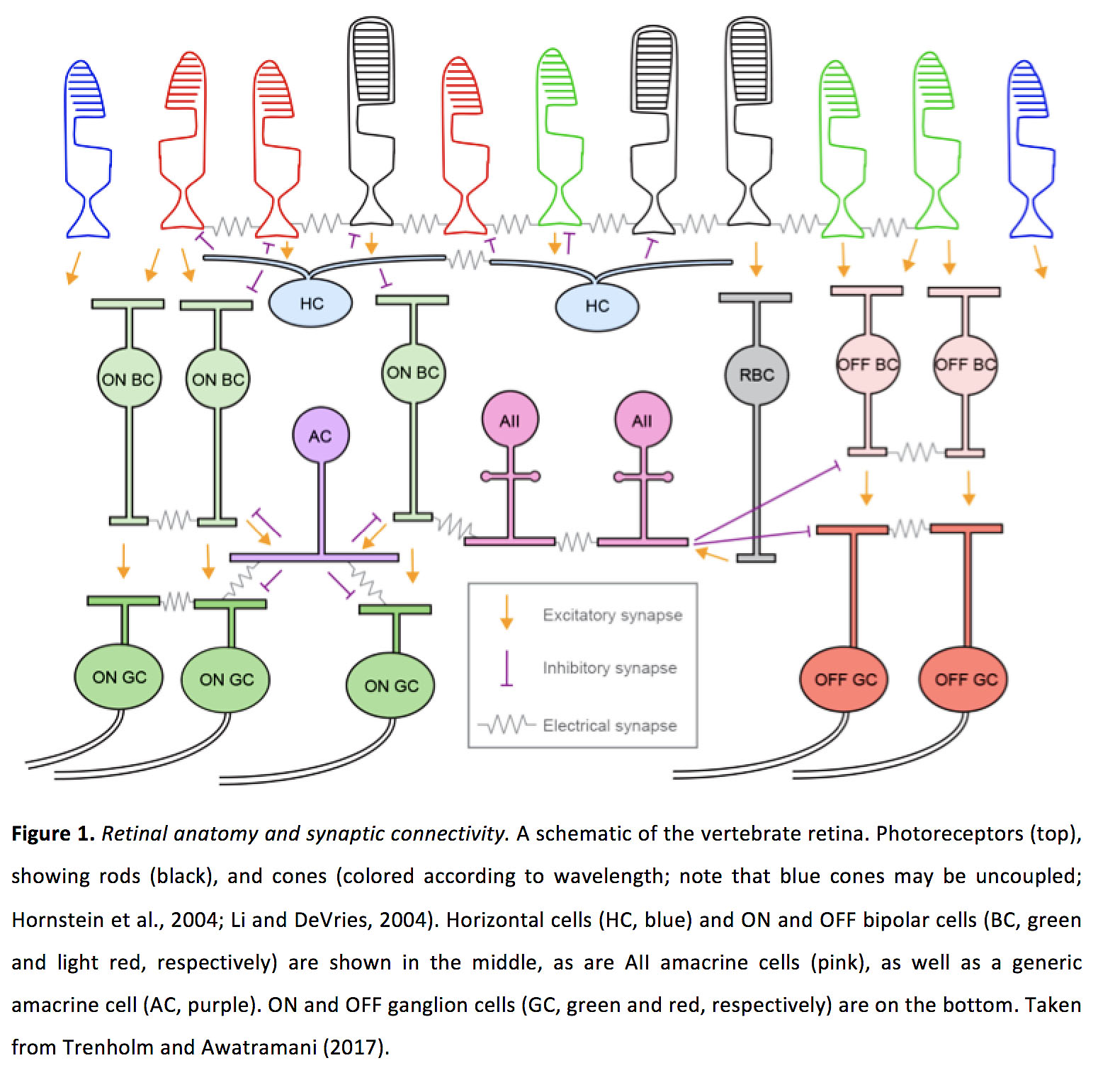
The vertebrate retina contains five principal neuron classes: photoreceptors (rods and cones), horizontal cells, bipolar cells, amacrine cells and ganglion cells. For each class of neuron, we describe electrical and chemical synaptic connectivity patterns (Figure 1). For electrical synapses, when known, we outline the connexin subtypes involved in forming gap junctions and describe their biophysical properties. Also, whenever possible, we describe the strength of gap junctions between pairs of neurons, represented as either the junctional conductance or the coupling coefficient (i.e. the change in membrane potential of a post-junctional cell divided by the change in the membrane potential in a pre-junctional cell when then membrane potential of the pre-junctional cell is altered).
Despite the characterizations of coupling strength that we will outline below, it is important to keep in mind that the conductance through a gap junction is not static, and changes to the conductance can have significant effects the roles that electrical synapses play in neuronal circuits. For instance, modifying the strength of gap junction coupling can significantly alter the receptive field size of retinal neurons by modifying the strength and extent of their excitatory receptive field surround. For many neurons in the retina, light adaptation or circadian rhythm can modulate the strength of electrical coupling and thus alter receptive field sizes. Changes to coupling strength could also modify other roles for gap junctions in retinal circuits. While molecular mechanisms underlying changes in coupling strength have been studied in detail (Ribelayga and O’Brien, 2017), in the following section we focus on the functional outcomes of modifications in coupling strength on receptive field size.
3. Photoreceptors
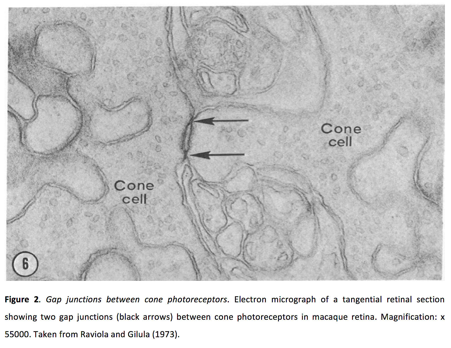
Vision starts in rod and cone photoreceptors, as photons are converted into an electro-chemical signal. Photoreceptors make glutamatergic synapses with horizontal and bipolar cells (ionotropic sign conserving synapses with horizontal cells and OFF bipolar cells; metabotropic sing-inverting synapses with ON bipolar cells). Photoreceptors also form both homologous (i.e. cone-cone and rod-rod) and heterologous (rod-cone) electrical synapses (Figure 2) (Baylor et al., 1971; Custer, 1973; Raviola and Gilula, 1973; Copenhagen and Owen, 1976; Tsukamoto et al., 2001; DeVries et al., 2002; Hornstein et al., 2004, 2005; Li et al., 2012).
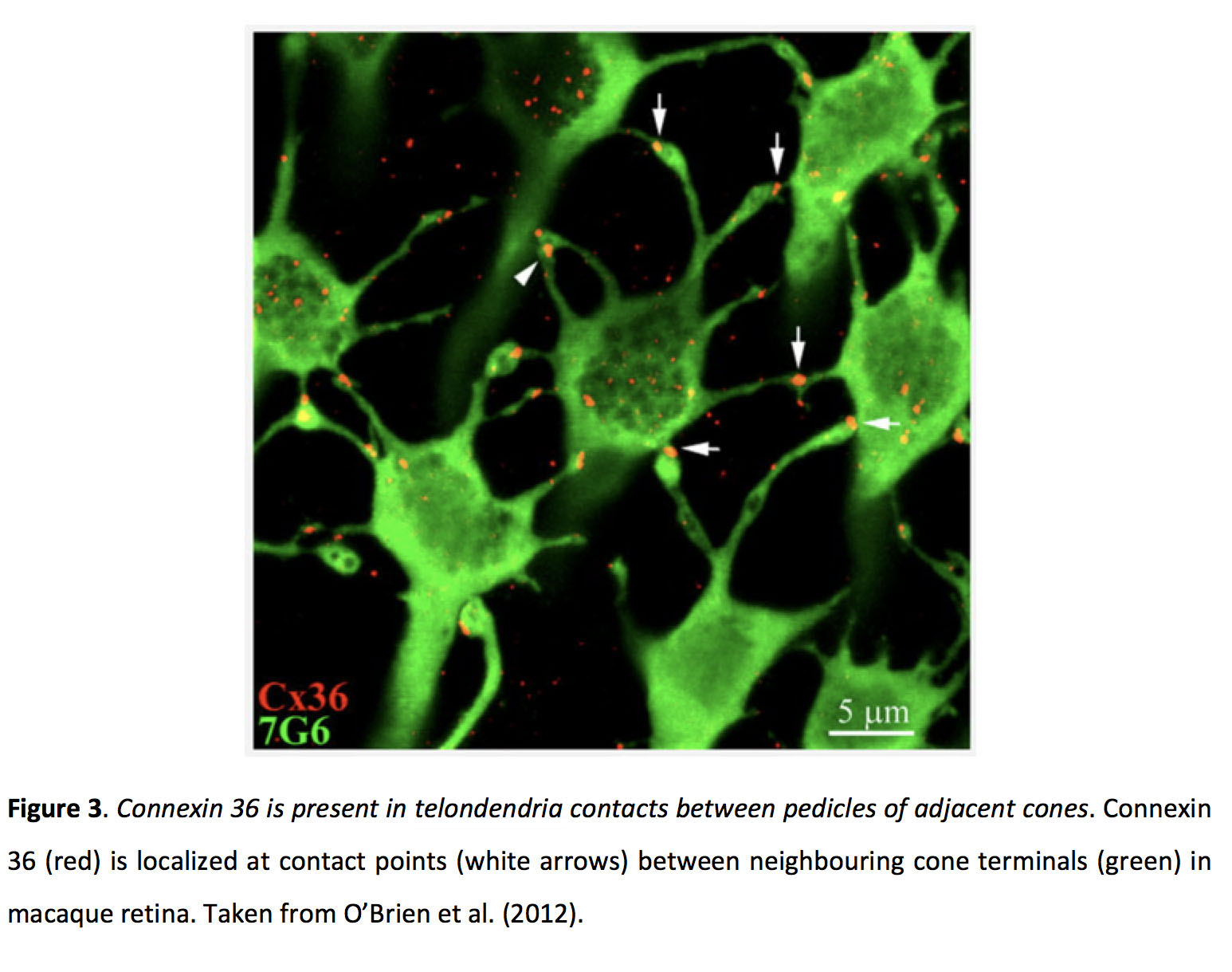
Cone gap junctions contain connexin 36 (Cx36) in mammals (Figure 3; Lee et al., 2003; Feigenspan et al., 2004; O’Brien et al., 2012; Kántor et al., 2016) and Cx35 in zebrafish (Li et al., 2009), while the connexin involved in rod electrical synapses has not been definitively identified (Lee et al., 2003; but see also Zhang and Wu, 2004; Li et al., 2013), and coupling between rods and cones appears to rely on Cx36 (Asteriti et al., 2017). Photoreceptor gap junctions are localized around the axon terminal of cones (Custer, 1973; Raviola and Gilula, 1973; Kolb, 1977; Tsukamoto et al., 1992, 2001; Li and DeVries, 2004).
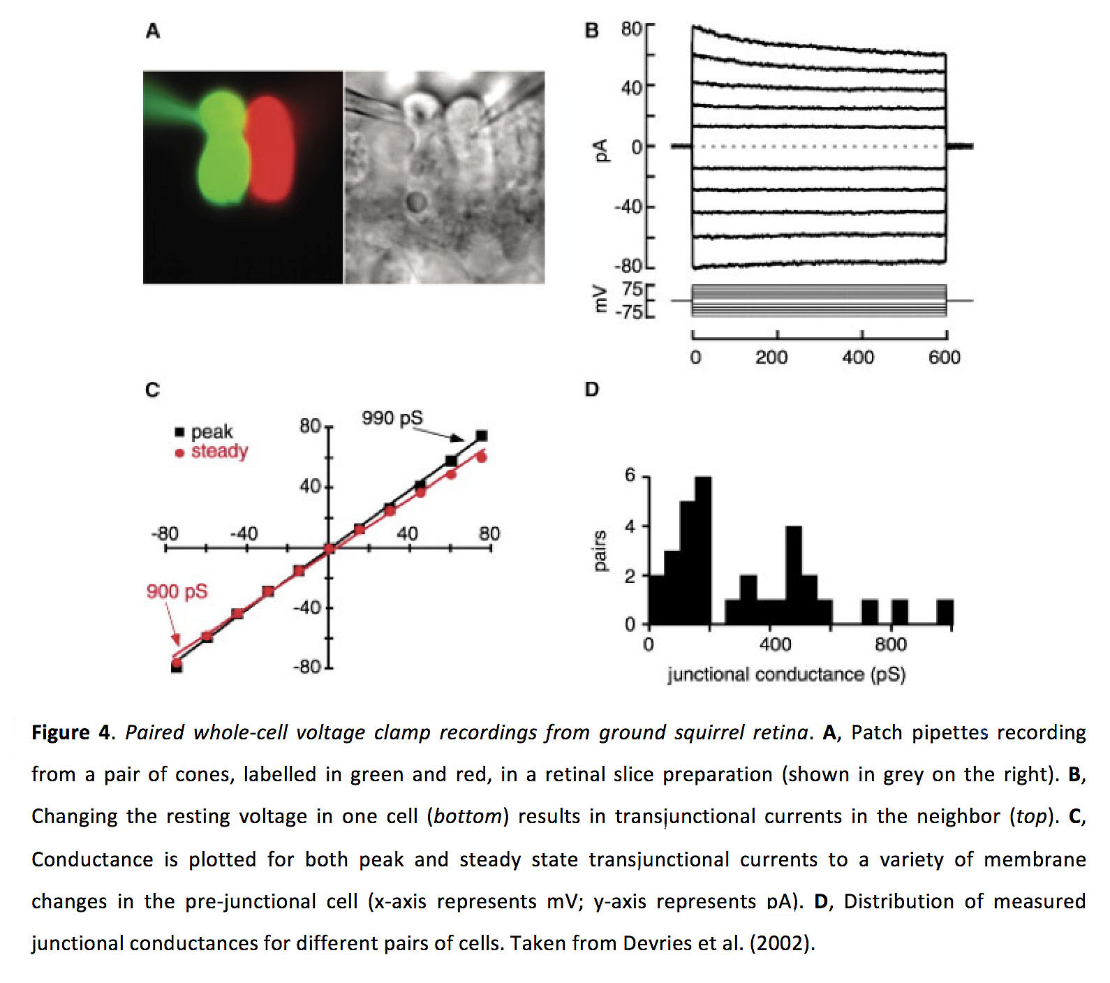
Based on physiological recordings, it appears that homologous photoreceptor coupling is stronger than heterologous coupling. The junctional conductance of cone-cone and rod-rod gap junctions appears to be bidirectionally symmetrical and in the range of 200-800 pS (Figure 4; DeVries et al., 2002; Hornstein et al., 2004; Li and DeVries, 2004; Zhang and Wu, 2005; Li et al., 2012), with a coupling coefficient of around 0.15 (Werblin, 1978; Hornstein et al., 2004). The junctional conductance of rod-cone gap junctions has been reported to be bidirectionally symmetrical and in the range of 40-200 pS (Gao et al., 2013), with a coupling coefficient of around 0.04 (Attwell et al., 1984). From tracer dye experiments where the dye is injected into cones, it appears that being coupled to rods is more common than being coupled to only cones or to both rods and cones, and some cones may be uncoupled (Hornstein et al., 2005). In contrast, from tracer dye experiments where the dye is injected into rods, rod-rod coupling seems to be more common than rod-cone coupling and some rods may be uncoupled (Li et al., 2012).
Circadian rhythm, and not light adaptation, appears to be the main controller for altering the strength of electrical coupling in photoreceptors in fish and mouse retinae (Ribelayga et al., 2008). Psychophysical experiments in humans have indicated that cone coupling appears to be unaffected by light adaptation, but effects of circadian rhythm have not been tested (DeVries et al., 2002). In contrast, light adaptation, not circadian rhythm, appears to modulate electrical coupling strength between salamander photoreceptors (Gao et al., 2013). Altering the strength of gap junctions can modify the receptive field size of photoreceptors. For instance, cones have larger receptive fields during states in which rods and cones are more strongly electrically coupled (Ribelayga et al., 2008). Similarly, rod receptive fields are also expected to be larger when they are more strongly coupled, and coupling decreases trial-to-trial variability in their responses to dim flashes (Li et al., 2012). Finally, heterotypic gap junctions allow cone responses to pass into rods (Wu and Yang, 1988), and rod responses to pass into cones (Figure 5; Nelson, 1977; Schneeweis and Schnapf, 1995; Hornstein et al., 2005; Asteriti et al., 2014, 2017), meaning that increases in electrical coupling should extend the range of light intensities over which a given rod or cone can respond, and at least for cones such coupling could result in increased reliability of threshold responses.

4. Horizontal cells
Horizontals receive glutamatergic inputs from photoreceptors, and in turn provide negative feedback to photoreceptors and feedforward inhibition to bipolar cells (though the nature of these interactions is still actively being studied; Thoreson and Mangel, 2012). Additionally, horizontal cells are densely interconnected with gap junctions (Figure 6; Raviola and Gilula, 1975; Kolb, 1977).

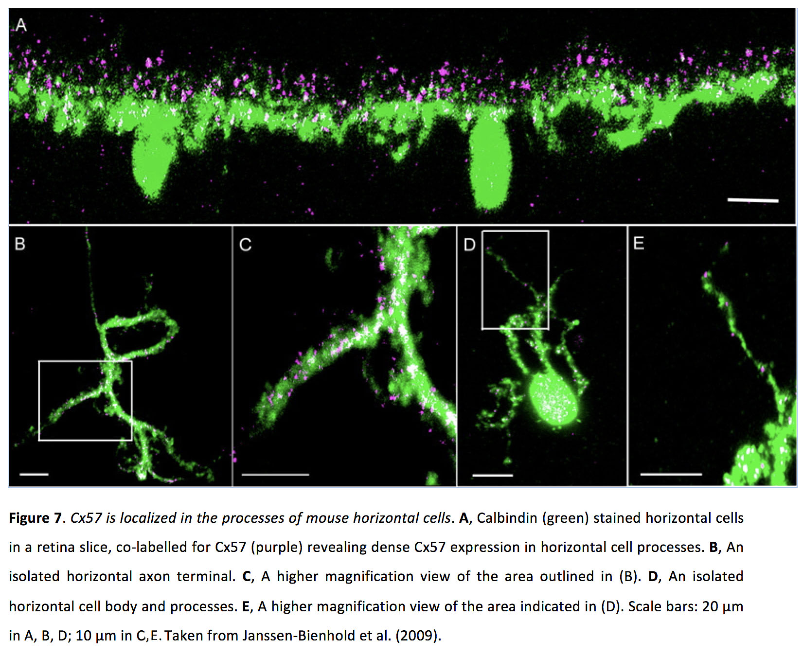
Horizontal cell gap junctions can be either axoaxonic and dendrodendritic. In mouse, Cx57 is present in both types of gap junctions (Figure 7; Hombach et al., 2004; Janssen-Bienhold et al., 2009), while Cx50 is localized in axonal gap junctions (Dorgau et al., 2015). In rabbit A-type horizontal cells, Cx50 localizes in dendritic gap junctions (O’Brien et al., 2006), and Cx57 is found in axonal gap junctions in B-type horizontal cells (Pan et al., 2012). In zebrafish, horizontal cell gap junctions appear to be comprised of Cx52.6, 52.9 and 55.5 (Shields et al., 2007; Klaassen et al., 2011).
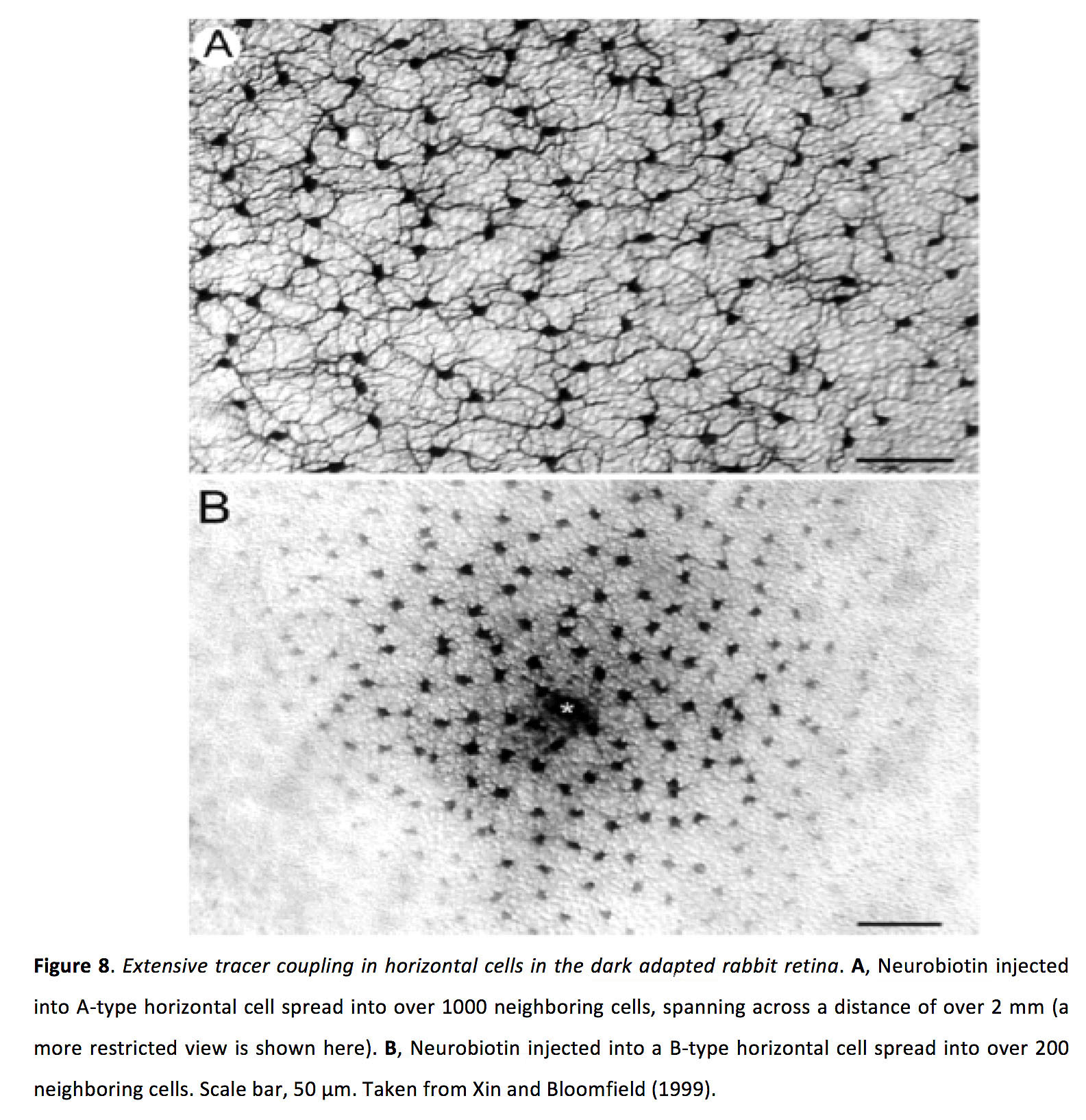
Coupling between horizontal cells appears to be very strong, with a bidirectional and symmetrical junctional conductance of > 1ns and with a coupling coefficient of 0.5 or greater (Lasater and Dowling, 1985; DeVries and Schwartz, 1989; McMahon, 1994; McMahon and Mattson, 1996; these recordings are from cultured horizontal cells). From remarkable tracer dye experiments, where dye is injected into a single horizontal cell, it has been found that dye can spread into more than 1000 surrounding horizontal cells extending across a distance of over 1 mm (Figure 8; Xin and Bloomfield, 1999).
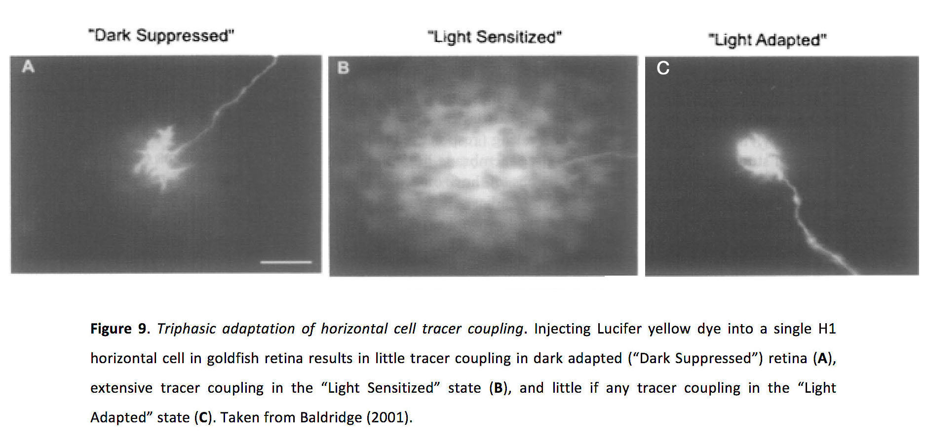
In horizontal cells, light adaptation appears to be the main factor controlling the strength of gap junction coupling (Ribelayga and Mangel, 2003, 2007). Horizontal cell coupling has been shown to exhibit a tri-phasic relationship depending on luminance levels: tracer coupling experiments show minimal coupling in the dark adapted or “dark-suppressed” state; tracer coupling is greatly increased in medium light levels, during the so-called “light-sensitized” state; tracer coupling is very weak during the light adapted state (Figure 9; Xin and Bloomfield, 1999; Baldridge, 2001).
Horizontal cell coupling seems to strongly affect receptive field size (Mangel and Dowling, 1985), with receptive fields being approximately three times larger in the light-sensitized state (Xin and Bloomfield, 1999). Consistent with this finding, knocking out Cx57 in mice decreases the size of horizontal cell receptive fields (Figure 10; Shelley et al., 2006).

As horizontal cells provide negative feedback to photoreceptors, it has been posited that they might be responsible for generating inhibitory surround receptive fields in downstream ganglion cells (Werblin and Dowling, 1969), in which case modifying horizontal cell coupling strength would modify ganglion cell receptive field size. However, the effect of horizontal cell feedback on ganglion cell surround responses remains uncertain, and at least in some cases horizontal cells do not drive significant surround responses in ganglion cells (Farrow et al., 2013; Hoggarth et al., 2015; Drinnenberg et al., 2018). Additionally, despite reducing horizontal cell receptive field size, knocking out Cx57 has little effect on ganglion cell spatial tuning and on visual acuity in mice (Dedek et al., 2008). In contrast, horizontal cells might play a role in temporal tuning of ganglion cells. For instance, it has been suggested that strong electrical coupling might functionally remove horizontal cells from the retinal circuit, by effectively shunting their input current from photoreceptors. In such a situation, modifying coupling strength could switch the retina from a mode of short integration time in daylight conditions to a mode of long integration time in nightlight conditions (Pandarinath et al., 2010). In another study, horizontal cell feedback to photoreceptors was shown to differentially affect transient and/or sustained portions of ganglion cell responses (Drinnenberg et al., 2018), and this could provide another mechanism whereby changes in horizontal cell coupling could affect temporal tuning in ganglion cells.
5. Bipolar cells
Bipolar cells, of which there are at least 10 types (Wässle et al., 2009), pass photoreceptor signals onto amacrine and ganglion cells at glutamatergic synapses. Additionally, bipolar cells can be electrically coupled to other bipolar cells and amacrine cells. Most commonly, bipolar cells are coupled to AII amacrine cells (Figure 11; Kolb and Famiglietti, 1974), though recent work shows that certain ON cone bipolar cells form gap junctions with non-AII amacrine cells (Farrow et al., 2013; Lee et al., 2015).

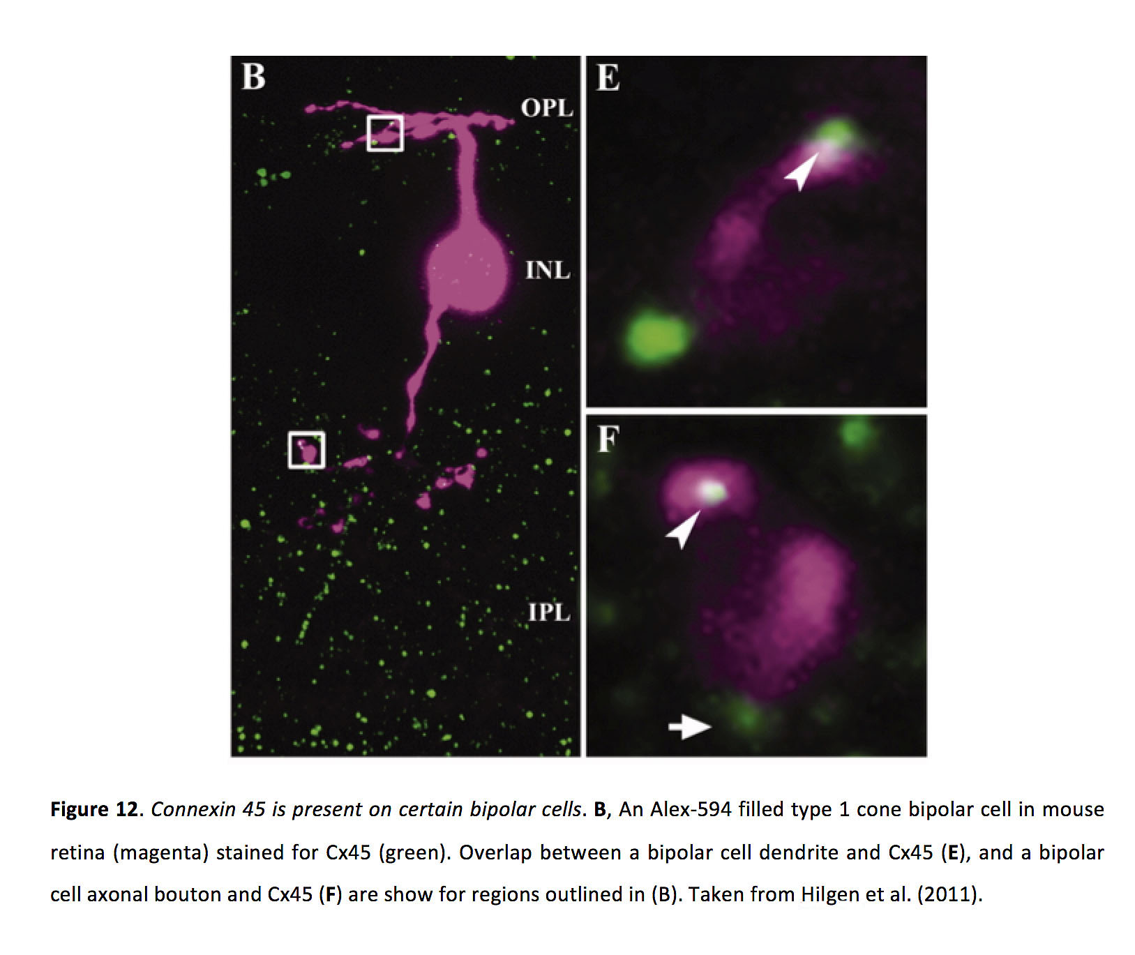
Gap junctions between bipolar cells can be localized in dendrites (Arai et al., 2010) or axon terminals (Jacoby and Marshak, 2000; Marc et al., 1988), though gap junctions between ON cone bipolar cells and AII amacrine cells are localized in the bipolar cell axon terminal (Strettoi et al., 1992). In mouse retina, bipolar cell gap junctions contain either Cx36 or Cx45 (Figure 12; Deans et al., 2002; Feigenspan et al., 2004; Han and Massey, 2005; Maxeiner et al., 2005; Hilgen et al., 2011).
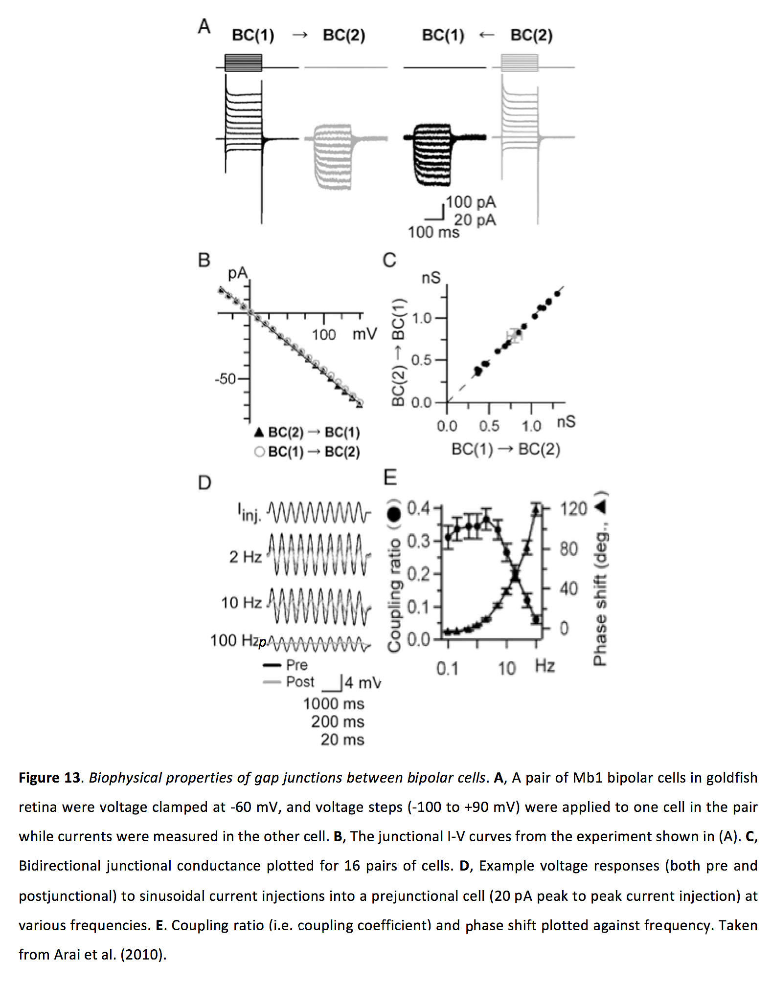
Bipolar cell to bipolar cell gap junctions are bidirectional and symmetrical, with a junctional conductance of around 1 nS and a coupling coefficient of around 0.3 (Figure 13; Kujiraoka and Saito, 1986; Umino et al., 1994; Mills, 1999; Dacey et al., 2000; Arai et al., 2010).
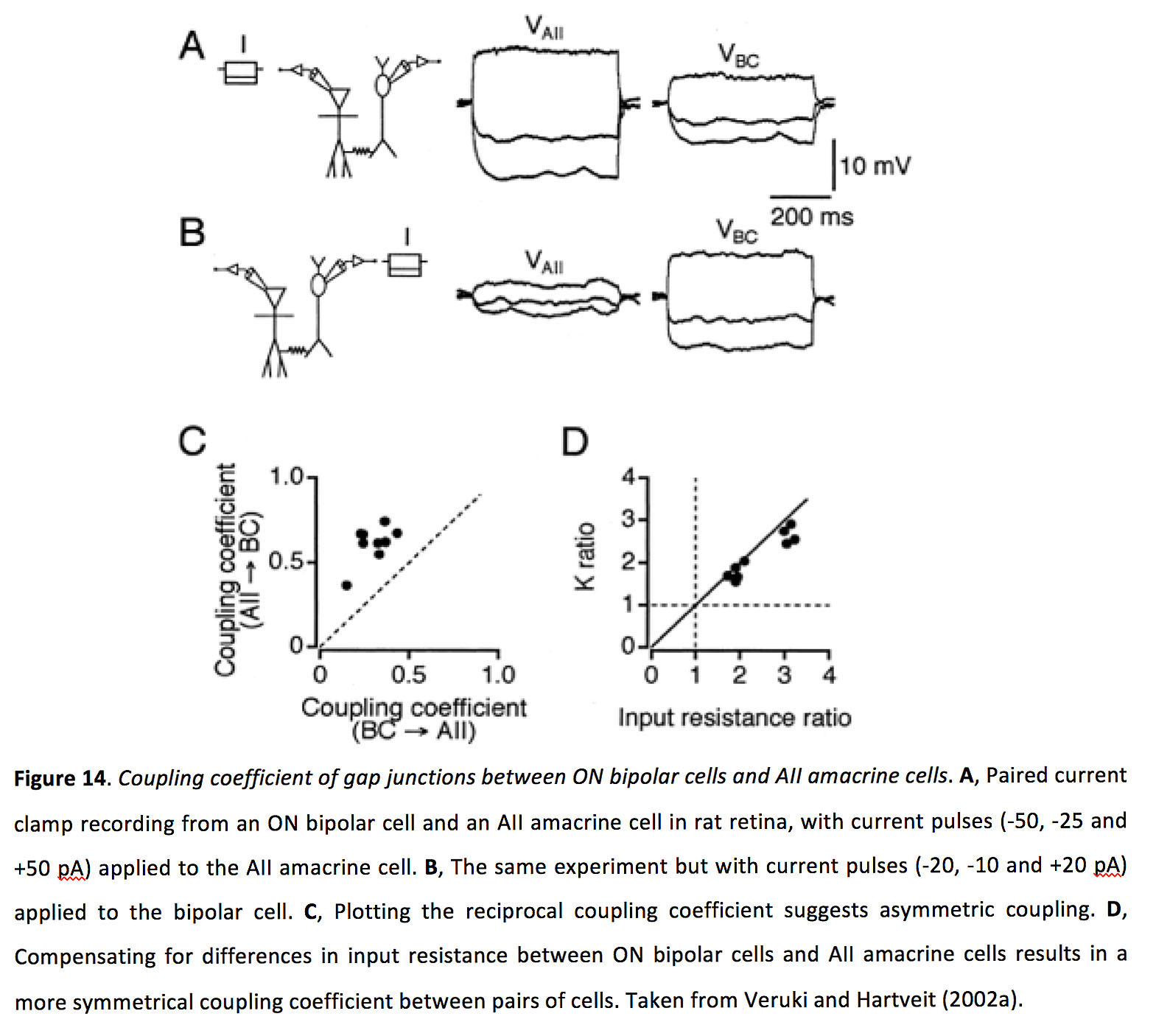
ON cone bipolar cells form gap junctions with AII amacrine cells that are bidirectional but asymmetric, with a junctional conductance of approximately 1 nS and a coupling coefficient between 0.3-0.6 (Kolb and Famiglietti, 1974; Deans et al., 2002; Veruki and Hartveit, 2002a; Marc et al., 2014; Graydon et al., 2018).
The gap junction conductance between electrically connected bipolar cells increases in the light adapted state (at least as shown in goldfish for Mb-1 bipolar cells; Arai et al., 2010). While it has been suggested that bipolar cell coupling can result in increased receptive field sizes, which some studies have found to be significantly larger than dendritic field sizes (Kujiraoka and Saito, 1986; Saito and Kujiraoka, 1988; Dacey et al., 2000; but see also Berntson and Taylor, 2000), to our knowledge the effect of changing light adaptation state on bipolar cell coupling and receptive field size has never been directly tested. Additionally, previous work has shown that antagonistic surround receptive fields in bipolar cells can appear only in the light adapted state (Werblin, 1970), which would be expected to counteract any excitatory surround receptive field properties provided via gap junction inputs from neighbouring cells. As such, while bipolar cell gap junctions appear to enhance their ability to integrate spatially separated stimuli and preferentially enhance their signaling of low contrast stimuli (Kuo et al., 2016), further study is required to define the effect of light adaptation on the strength of gap junction coupling for bipolar cells, and for examining whether such changes effectively modify receptive field size.
6. Amacrine cells
Amacrine cells, of which there are more than 45 types (Masland, 2012b; Diamond, 2017), provide inhibition, via GABA or glycine release, to bipolar cells, other amacrine cells and ganglion cells. Some amacrine cells can also co-release acetylcholine (Masland and Mills, 1979; Lee et al., 2010; Sethuramanujam et al., 2016), dopamine (Zhang et al., 2007), or glutamate (Baden and Euler, 2016). Many amacrine cells form homologous gap junctions (Xin and Bloomfield, 1997; Li et al., 2002; Veruki and Hartveit, 2002b; Abdel-Majid et al., 2005; Marc et al., 2014), and heterologous gap junctions with bipolar cells – as described above – and ganglion cells (Xin and Bloomfield, 1997; Völgyi et al., 2009). AII amacrine cells form gap junctions with one another in their dendritic arbors (Figure 15; Strettoi et al., 1992). It seems likely that other amacrine cell types also make dendrodendritic gap junctions with amacrine and ganglion cells, as this is often the only location where their processes overlap.

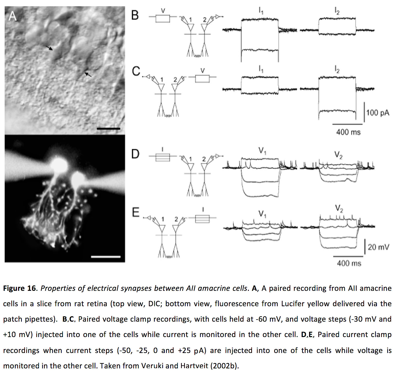
AII amacrine cells appear to form gap junctions with ON cone bipolar cells with Cx36. Coupling between AII amacrine cells is bidirectional and symmetric, with a junctional conductance of ~700 pS and a coupling coefficient of ~ 0.3 (Figure 16; Veruki and Hartveit, 2002b).
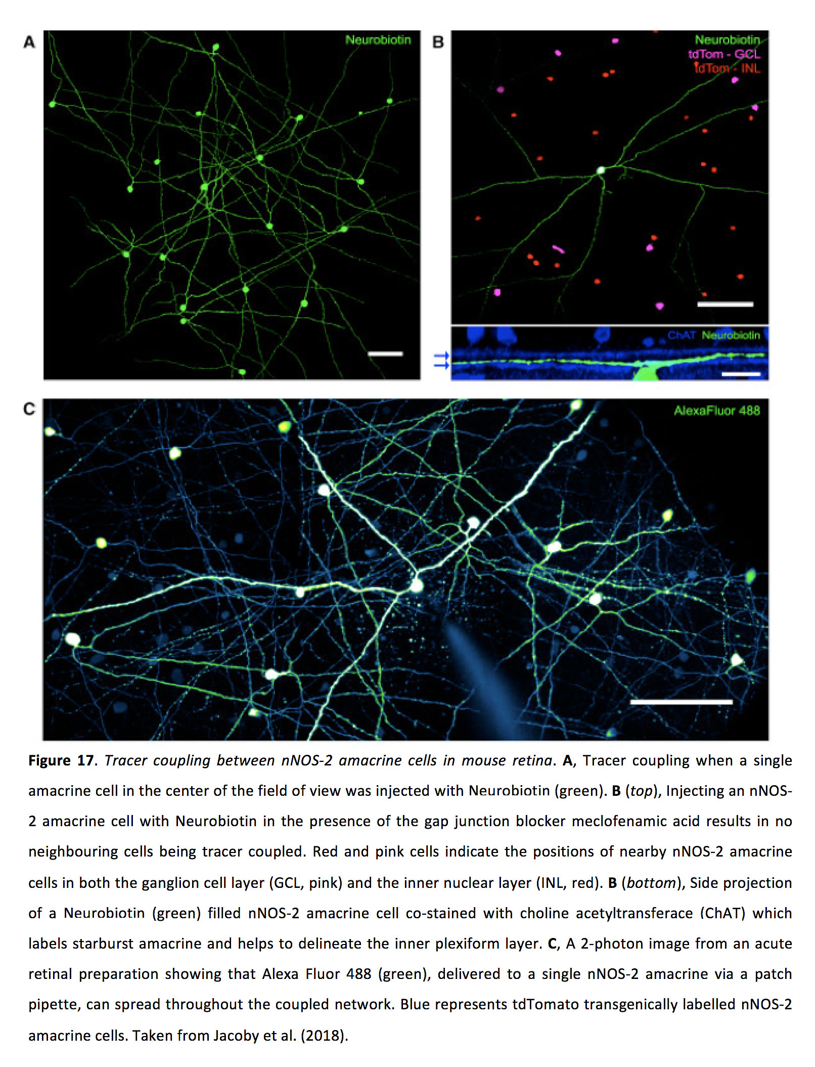
Other amacrine cell types form gap junctions with amacrine or ganglion cells with electrical synapses comprising either Cx36 or Cx45 (Pan et al., 2010; Pang et al., 2013). nNOS-2 amacrine cells, which are the main source of nitric oxide in the mouse retina, have been shown be extensively interconnected with what appear to be Cx45 containing gap junctions, and exhibit a bidirectional and symmetrical conductance, with a junctional conductance that was modeled to be very large (4.4 nS; Figure 17; Jacoby et al., 2018). The measured coupling coefficient for nNOS-2 amacrine cell gap junctions was ~0.08 (Jacoby et al., 2018).
Light adaptation state appears to strongly control coupling strength in amacrine cells (Bloomfield and Xin, 1997). Similar to horizontal cells, tracer coupling experiments indicate that AII amacrine cells exhibit minimal coupling during both very dim and bright conditions, and strong coupling during mid light conditions (Figure 18; Bloomfield et al., 1997).

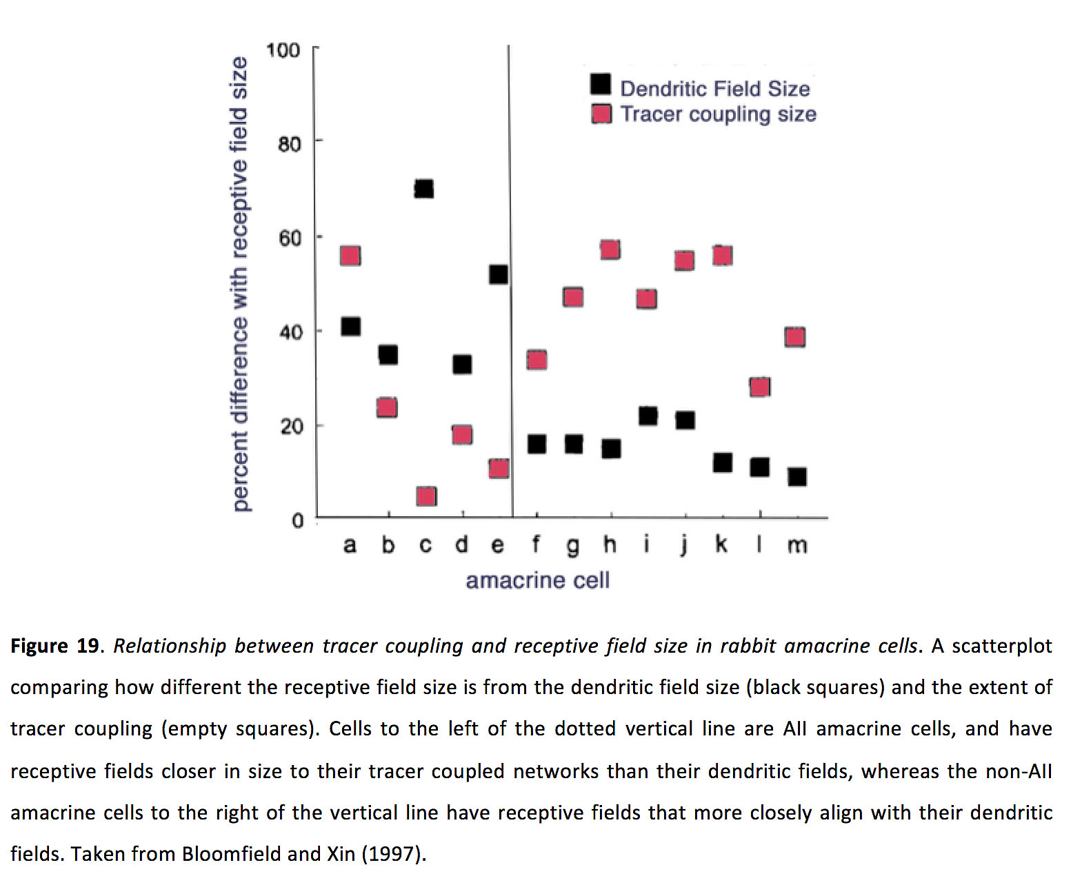
Such changes in coupling strength appear to strongly influence receptive field size, as AII amacrine cell receptive fields can be 2-3 times larger than their dendritic fields in the light-sensitized state (Bloomfield and Xin, 1997). However, a direct relationship between coupling strength and receptive field size does not appear to hold across all amacrine cell types. For instance, unlike AII amacrine cells, work with some other types of amacrine cells did not indicate a strong effect of light adaptation on receptive field size, despite the fact that tracer coupling experiments showed light-adaptation dependent changes in tracer coupling patterns (Figure 19; Bloomfield and Xin, 1997). Why the effect of gap junction coupling on receptive field size does not appear consistent across all amacrine cell types remains unclear, though it could have to do with the fact that the dendrites of some amacrine cell types appear to perform local computations (Grimes et al., 2010), which might therefore not require global changes in receptive field size for changes in junctional conductance to have an effect on circuit function. Another possibility for the difference of light adaptation on amacrine cell coupling could be differential regulation by dopamine. Indeed, gap junction coupling of A8 amacrine cells, which are coupled to both ON and OFF bipolar cells with Cx36 gap junctions, does not appear to be modulated by dopamine receptor D1 activation, in contrast to AII amacrine cells (Yadav et al., 2019). Finally, in mouse nNOS-2 amacrine cells, there appears to be a remarkable self-regulation between light activation and coupling state. nNOS-2 cells are extensively electrically coupled in the dark adapted retina, whereas upon light activation they produce nitric oxide which in turn decouples the network (Jacoby et al., 2018). Whether this light-activated decoupling affects receptive field size remained to be tested.
7. Ganglion cells
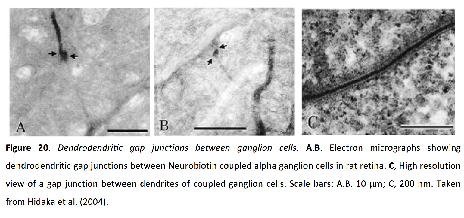
Ganglion cells, of which there are upwards of 30 types (Dacey, 2004; Sanes and Masland, 2015; Baden et al., 2016), receive excitatory glutamatergic input from bipolar cells and inhibitory GABAergic and glycinergic input from amacrine cells, and send their signals, via the optic nerve, to multiple higher visual areas (Robles et al., 2014; Martersteck et al., 2017). Ganglion cells also form dendrodendritic gap junctions with amacrine cells and other ganglion cells (Figure 20; Vaney, 1991, 1994; Xin and Bloomfield, 1997; Hidaka et al., 2004; Völgyi et al., 2009; Müller et al., 2010a; Reifler et al., 2015).
.
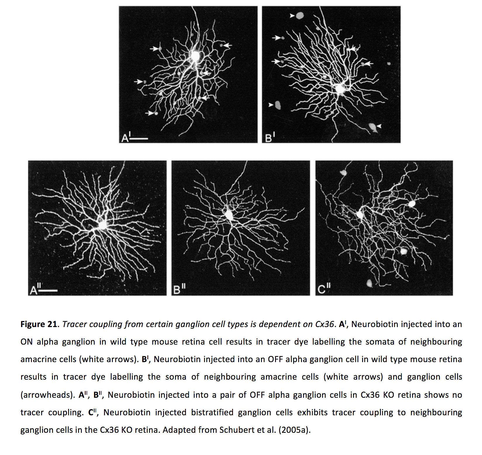
Ganglion cell gap junctions are frequently comprised of connexin 36 (Figure 21; Schubert et al., 2005a; Pan et al., 2010; Yao et al., 2018; Kántor et al., 2018), though some contain connexin 45 (Schubert et al., 2005b) and possibly also connexin 30.2 (Müller et al., 2010b).
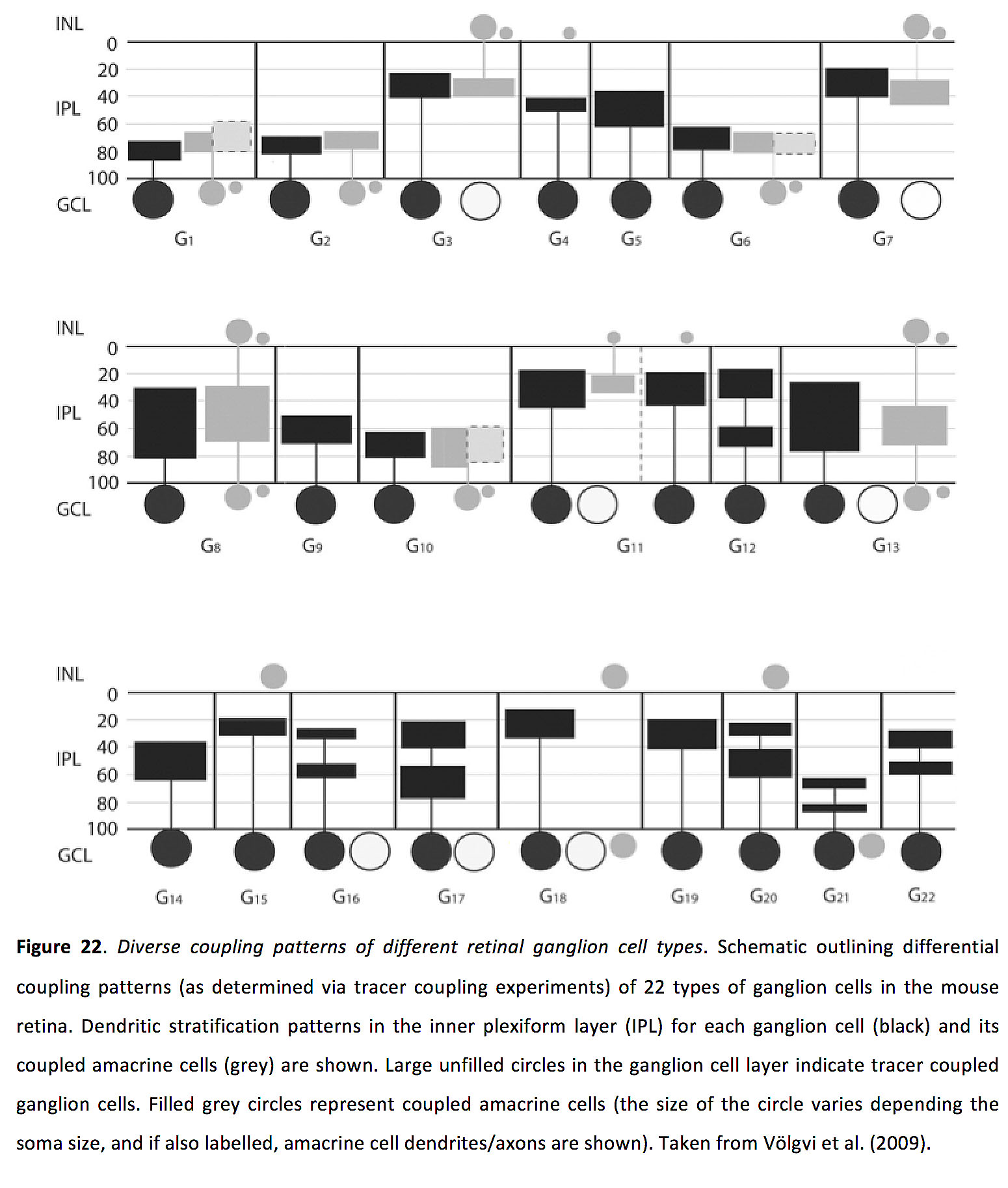
 Ganglion cell-ganglion cell coupling appears to be exclusively homologous, with no coupling between different ganglion cell types (Vaney, 1994; Hu and Bloomfield, 2003; Völgyi et al., 2009; Trenholm et al., 2013a), and distinct ganglion cell subtypes have unique coupling patterns (Figure 22; Völgyi et al., 2009). Less is known about ganglion cell-amacrine cell coupling, though tracer coupling work suggests that specific ganglion cells form gap junctions with specific types of amacrine cells (Figure 22; Völgyi et al., 2009).
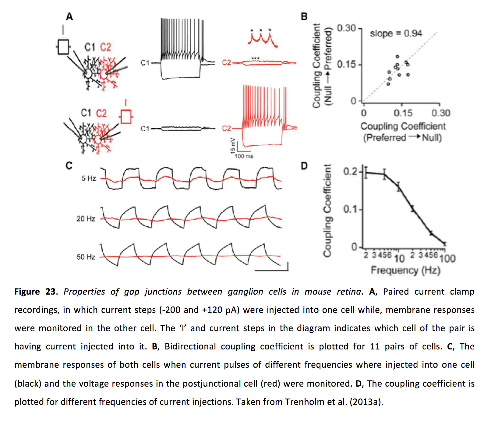
 Gap junctions between ganglion cells are bidirectional and have a symmetrical junctional conductance of around 1 nS and a coupling coefficient of around 0.15 (Figure 23; Hidaka et al., 2004; Trenholm et al., 2013a).
In ganglion cells, light adaptation appears to control coupling strength, with coupling being reduced in the dark adapted state and increased in the light adapted state (at least as has been shown for OFF α cells in the rabbit retina; Hu et al., 2010; though this may not be the case for all ganglion cell types (Yao et al., 2018)). Interestingly, while increased coupling strength does not seem to alter the size of the classical (spiking) receptive field (Hu et al., 2010; but see also DeVries, 1999), gap junction inputs from neighboring cells endow electrically coupled ganglion cells with extensive subthreshold excitatory surround receptive fields (Trenholm et al., 2013a). Furthermore, for many ganglion cells, their receptive field size actually decreases in light adapted conditions (Barlow et al., 1957; Peichl and Wässle, 1983; Muller and Dacheux, 1997), likely due to a strengthening of inhibitory surround responses arising from either horizontal and/or amacrine cells. For instance, it was found that for mouse ON and OFF α-like ganglion cells (Farrow et al., 2013) and directionally selective ganglion cells (Hoggarth et al., 2015), surround inhibition in ganglion cells is provided by wide-field amacrine cells, and this surround inhibition appears to be activated only at light intensities that activate cones. In this bright light, amacrine cell mediated surround inhibition is thought to rely on electrical coupling between a wide-field amacrine and ON cone bipolar cells. Knocking out Cx36 eliminates the bright light induced reduction in receptive field size of α-like ganglion cells (Farrow et al., 2013). As such, classical receptive field size in ganglion cells appears to be most strongly controlled by amacrine cell mediated surround inhibition (one recent study suggests that horizontal cells may only contribute as little as ~15% to the surround inhibition of ganglion cells (Drinnenberg et al. (2018)).
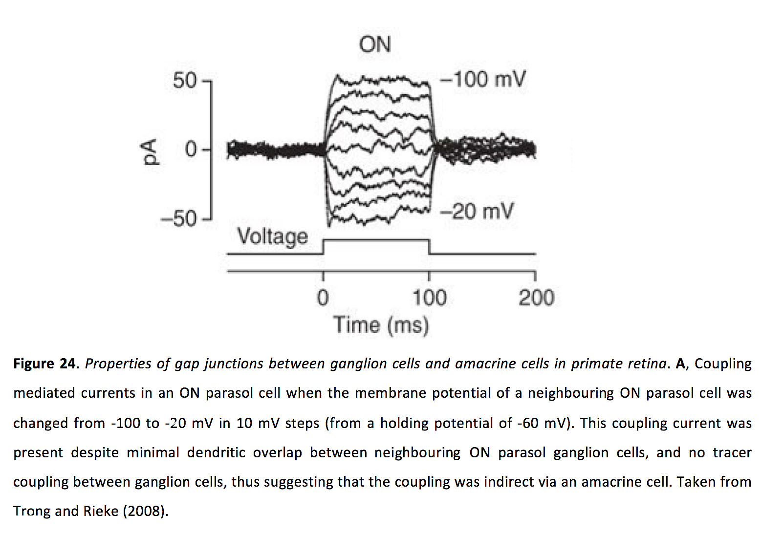
Much less is known about the properties of ganglion cell-amacrine cell coupling. Nonetheless, the gap junction strength appears to be strong enough such that current injected into one ganglion cell can pass via gap junctions into an amacrine cell, and then via additional gap junctions into a nearby ganglion cell, driving measurable responses in the indirectly coupled ganglion cell (Figure 24; Trong and Rieke, 2008).
8. Gap junctions and scotopic vision
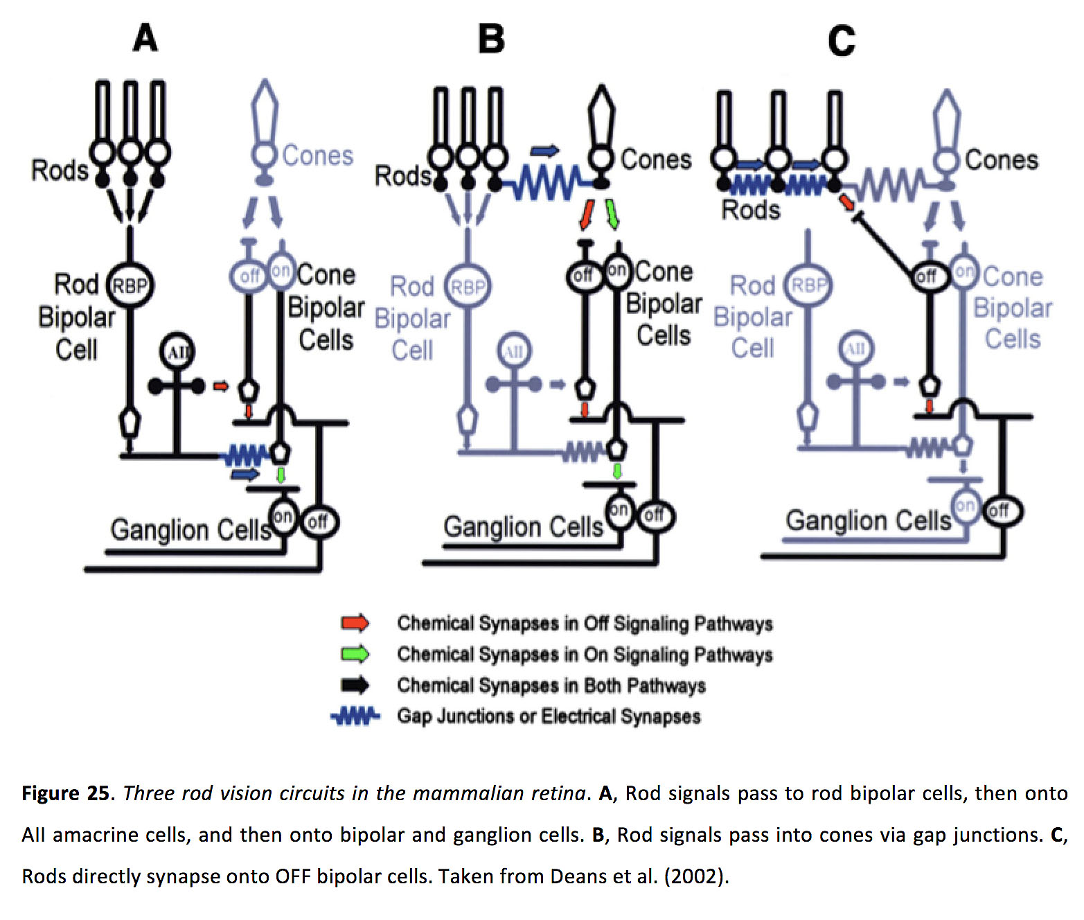
In dark (scotopic) light conditions, when rods are the active photoreceptor, gap junctions are critical for passing rod-mediated signals throughout the retina. In the classical rod pathway, rods form glutamatergic synapses with rod bipolar cells, which in turn form glutamatergic synapses with AII amacrine. These AII amacrine cells then transfer rod-generated signals to the ON cone bipolar cells, via gap junctions with ON cone bipolar cells. In addition AII amacrine cells also make glycinergic synapses with OFF bipolar cells (Figure 25; Kolb and Famiglietti, 1974; Völgyi et al., 2004; Ivanova et al., 2006; Münch et al., 2009; Marc et al., 2014; Graydon et al., 2018). In the second route, rod signals pass directly into cones, via rod-cone electrical synapses, and .thus passed onto the inner retina (Figure 25; Völgyi et al., 2004; Trexler et al., 2005; Abd-El-Barr et al., 2009). In the third route, some rods appear to form glutamatergic synapses with OFF bipolar cells, and pooling of signals between electrically coupled rods has been hypothesized to increase sensitivity of this pathway (Figure 25; Soucy et al., 1998; Tsukamoto et al., 2001; Li et al., 2004).
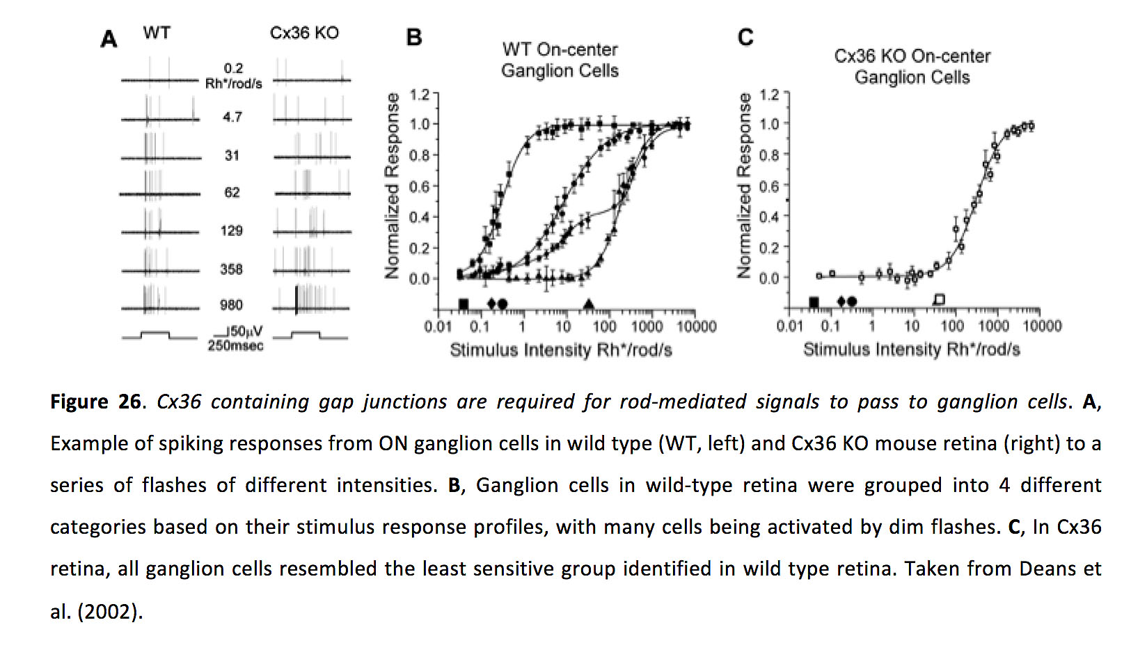
Consistent with an important role for gap junction signaling during rod-mediated scotopic vision, knocking out Cx36 greatly impairs rod-mediated responses in the inner retina (Figure 26; Deans et al., 2002; Völgyi et al., 2004; Abd-El-Barr et al., 2009). Additionally, gap junctions between certain types of amacrine and ganglion cells could also aid in high-sensitivity detection of dim light (Murphy and Rieke, 2011).
Gap junctions are thought to play another important role in rod signaling by allowing rods to increase their signal-to-noise ratio. Intrinsic noise in photoreceptors is unique to individual cells, whereas gap junctional coupling allows light-evoked signals to be shared, resulting in more effective detection of relatively weak signals and a decrease in response variability (note however that these gains may result in a loss of absolute sensitivity and resolution in dim conditions; Fain, 1975; Lamb and Simon, 1976; Attwell et al., 1984; Tessier-Lavigne and Attwell, 1988; DeVries et al., 2002; Hornstein et al., 2005; Li et al., 2012). A final possible role for gap junctions in rod signaling relates to the nature of synaptic transmission between rods and rod bipolar cells. There appears to be significant signal clipping (i.e. saturation of synaptic transmission) between rods and rod bipolar cells, and it has been posited that electrical coupling between rods could allow spatially-restricted visual stimuli to activate several rods, via junctional spread, and that this activated group of rods would then more effectively activate bipolar cells (Attwell et al., 1987).
9. Gap junctions and synchrony
One of the best described roles for gap junctions in the CNS is that they can synchronize activity between coupled cells (Connors and Long, 2004; Pereda et al., 2013). Classical work in the retina revealed that neighboring retinal ganglion cells exhibit significant spike synchrony (Arnett and Spraker, 1981; Mastronarde, 1983a, 1983b, 1983c). Subsequent studies verified that electrical synapses are responsible for driving diverse types of fine-scale synchrony between ganglion cells, across species (Brivanlou et al., 1998; DeVries, 1999; Hu and Bloomfield, 2003; Greschner et al., 2011; Völgyi et al., 2013; Trenholm et al., 2014).
Correlations between neighbouring pairs of ganglion cells are generally classified, based on temporal characteristics, as either narrow, medium or broad (Figure 27; Mastronarde, 1989; Brivanlou et al., 1998; Völgyi et al., 2013). Narrow synchrony occurs within ± 2 ms, with a trough in the cross-correlogram at 0 ms delay. Medium correlations have a cross-correlogram peak at 0 ms delay, and occur somewhere between 2-10 ms (Mastronarde, 1989), ~ 25-50 ms (Brivanlou et al., 1998), and less than 100 ms (Völgyi et al., 2013). Broad correlations generally exhibit a 0 ms delay peak in the cross-correlogram, and occur on a timescale of around 50 ms (Mastronarde, 1989), greater than 50 ms (Brivanlou et al., 1998) and greater than 100 ms (Völgyi et al., 2013), though between pairs of ON and OFF ganglion cells, broad correlations can exhibit a trough at 0 ms, indicative of a slow anti-correlation (Mastronarde, 1983a, 1983b, 1989).
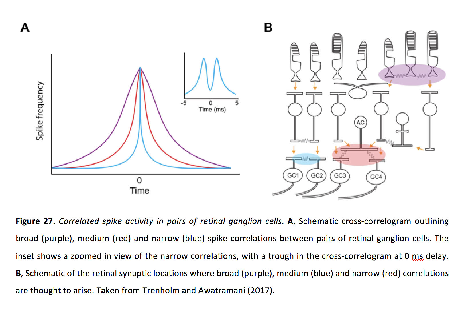
Pharmacological and genetic knockout experiments have been performed to tease apart the role of electrical and chemical synaptic transmission in these different types of ganglion cell correlations. Pharmacological studies indicate that narrow and medium correlations are unaffected by a cocktail of chemical synaptic blockers, but are inhibited by application of gap junction blockers (Brivanlou et al., 1998). Cx36 knockout experiments have indicated that broad correlations may also rely upon gap junction signaling (Völgyi et al., 2013). Based on these studies, it is thought that broad correlations arise initially in photoreceptors. In contrast, medium correlations are thought to be generated when pairs of ganglion cells are indirectly coupled via an amacrine cell (Mastronarde, 1989; DeVries, 1999; Brivanlou et al., 1998; Schnitzer and Meister, 2003; Völgyi et al., 2013), and narrow correlations form between reciprocally coupled ganglion cells (Mastronarde, 1983c; Brivanlou et al., 1998; DeVries, 1999; Hu and Bloomfield, 2003; Hidaka et al., 2004; Völgyi et al., 2013; Trenholm et al., 2014; Yao et al., 2018). In the rest of this section, we mostly focus on homologous coupling between ganglion cells and narrow correlations.
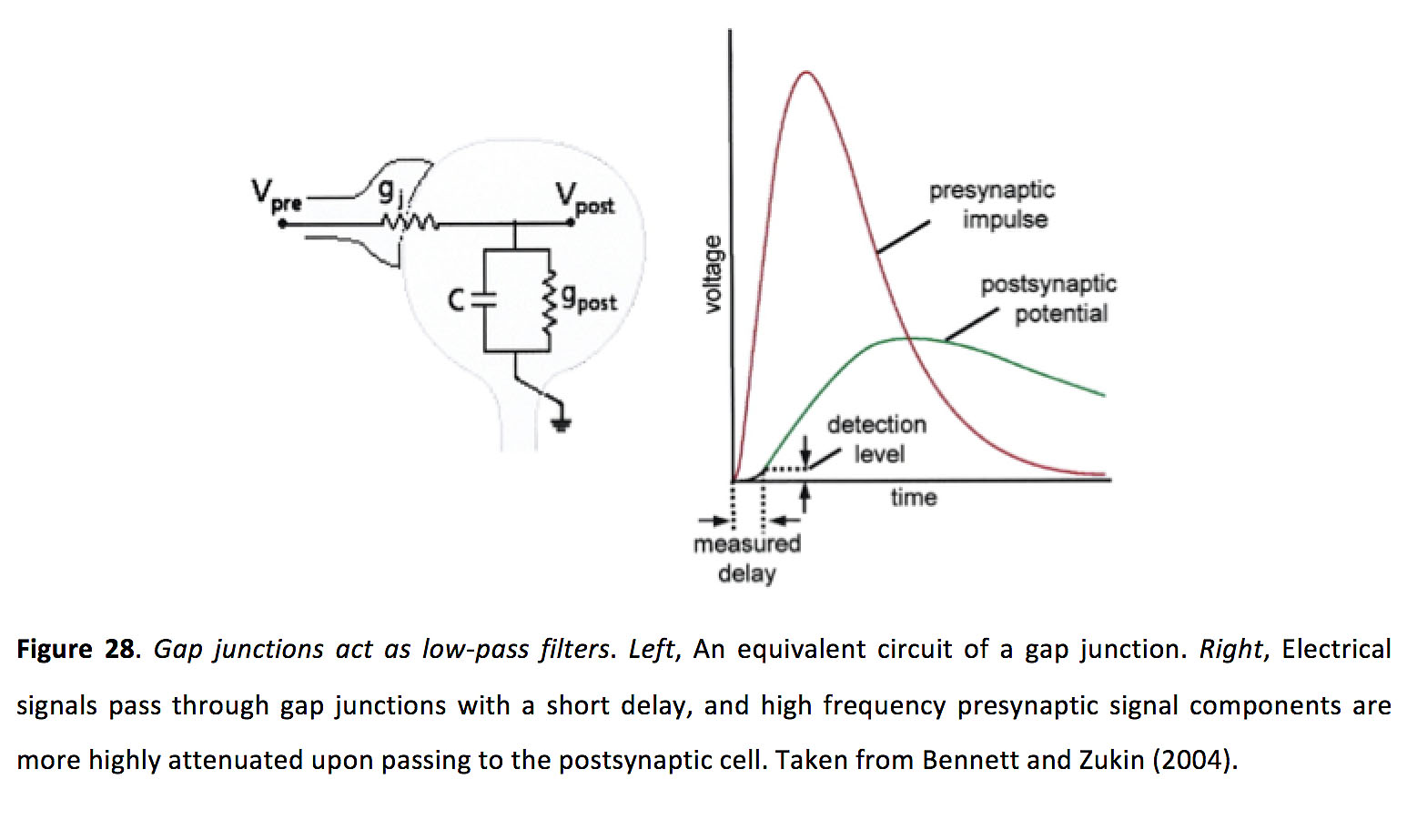
It is thought that narrow spike synchrony arises when one neuron fires an action potential and thus biases spike firing in its neighbor within a short time window. However, as gap junctions act as low-pass filters which significantly reduce the amplitude of action potential mediated signals as they pass across the junction (Figure 28; Bennett and Zukin, 2004; Connors and Long, 2004; Pereda et al., 2013), it was not immediately evident how such small signals can generate such robust synchrony (Trong and Rieke, 2008).
While it has been suggested that an action potential in one ganglion cell can directly drive a spike in a coupled neighbour (Hu and Bloomfield, 2003), more direct experimental evidence indicates that action potentials in one ganglion cell practically never drive suprathreshold responses in coupled ganglion cells (Trenholm et al., 2014). Consistent with this finding, early work in the field suggested that a spike in one ganglion cell will directly drive a spike in a coupled neighbor with a success rate of less than 5% (Mastronarde, 1983c). Furthermore, the classical receptive field of ganglion cells is not significantly increased by the presence of gap junctions (Hu et al., 2010; Trenholm et al., 2013; but see also DeVries, 1999). Why is a spike in one ganglion cell ineffective in directly driving spikes in a neighour? It turns out that in ganglion cells, coupled spikelets – at least when measured with a patch-pipette at the post-junctional soma – are small in amplitude (Figure 23; ~ 1 mV), though they do arrive with little delay (< 1 ms; Trenholm et al., 2013). As such, coupling between ganglion cells is similar to coupling between most other neurons in the CNS, in which spiking in a pre-junctional neuron only drives subthreshold activity in post-junctional neighbors (Connors and Long, 2004). How then do subthreshold coupled spikelets drive synchronous activity in coupled ganglion cells?
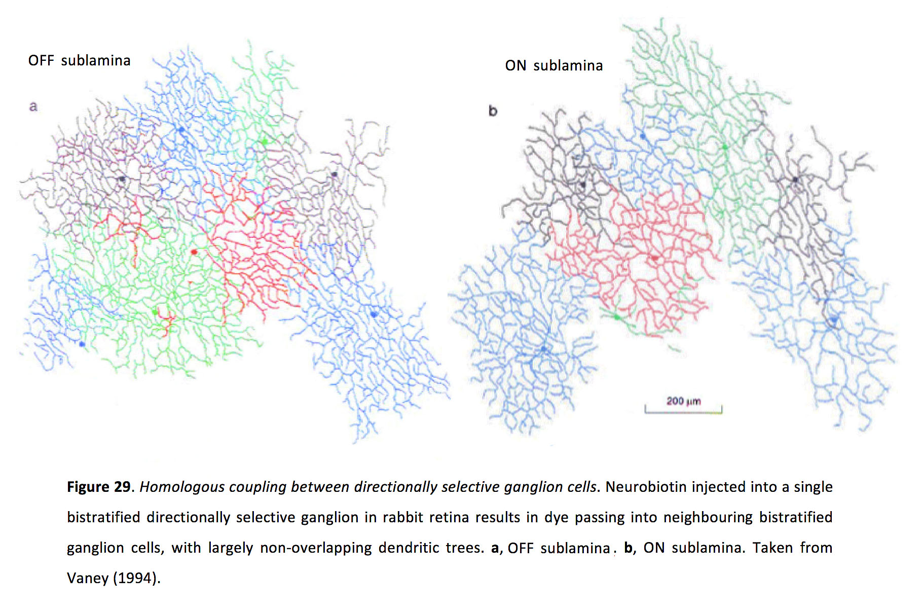
ON-OFF directionally selective (DS) ganglion cells have been an ideal model for studying how coupled spikelets can drive synchrony via non-linear dendritic signaling. DS ganglion cells generate Na+-channel dependent, TTX-sensitive dendritic spikes that trigger action potentials with high reliability (Oesch et al., 2005; Sivyer and Williams, 2013). There are at least four types of ON-OFF DS ganglion cells, coding the cardinal directions (Oyster and Barlow, 1967). For reasons that are not entirely clear, only ON-OFF DS cells that prefer superior motion in the visual field (ventral motion on the retina) exhibit homologous electrical coupling in the adult mammalian retina (at least in rabbit and mouse retina; Figure 29; Vaney, 1994; Trenholm et al., 2013; Xu et al., 2013).
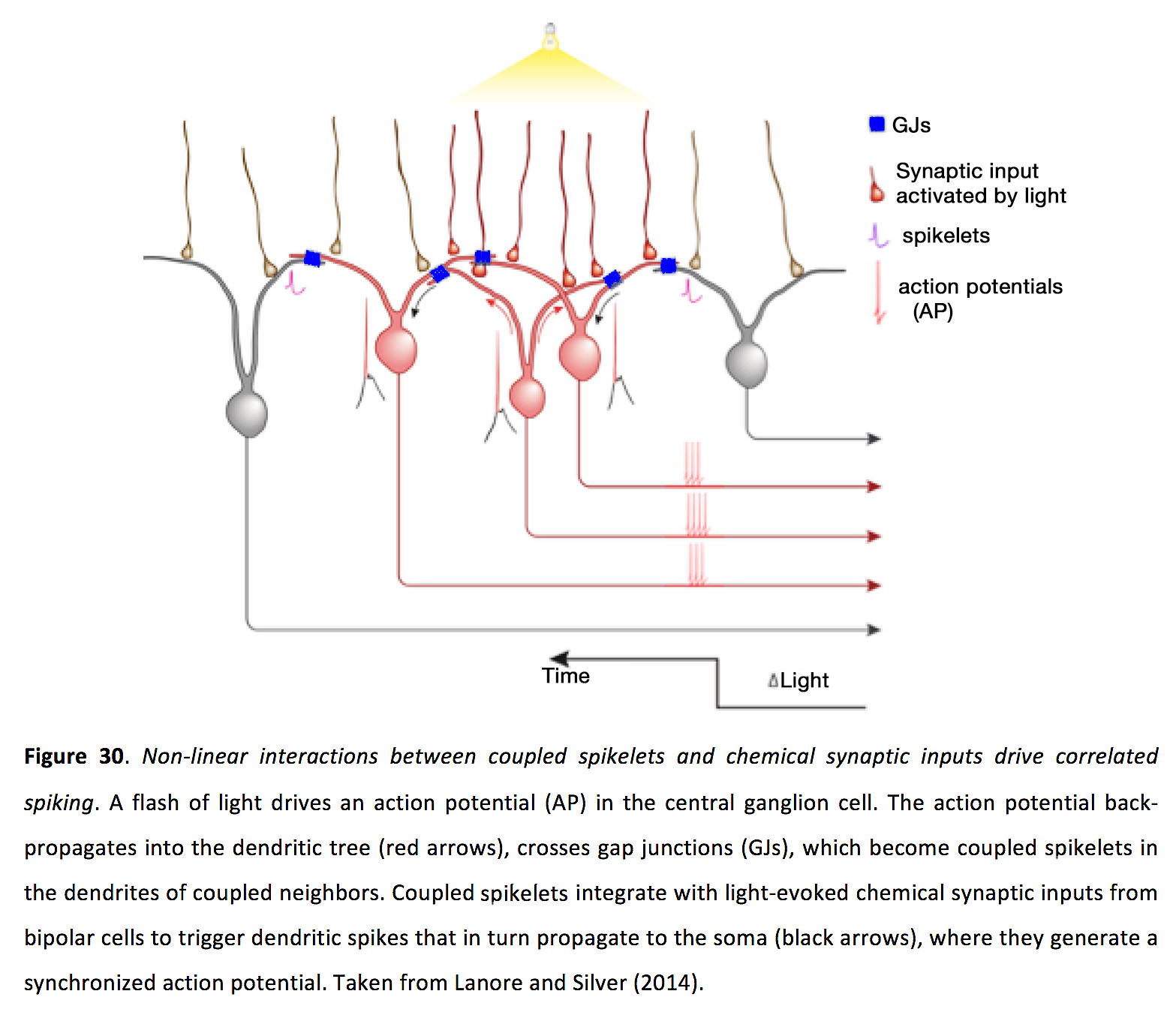
Interestingly, when coupled spikelets were simulated via current injection at the soma (with a patch pipette) in coupled DS ganglion cells, they did not significantly modulate action potential timing, raising the possibility that their site of action was in the dendrites (Trenholm et al., 2014). Furthermore, coupled spikelets do not appear large enough to directly initiate dendritic spikes themselves. It turns out that to drive spike synchrony in neighbouring cells, coupled spikelets must interact non-linearly with chemical synaptic inputs in the overlapping dendrites of neighbouring ganglion cells (Trenholm et al., 2014). Flashing light at a shared location of receptive overlap between a pair of neighboring coupled ganglion cells drives significant spike correlations, whereas flashing two spots of light at two different spatial locations to activate the same pair of ganglions at the same time but with non-overlapping chemical synaptic inputs, does not drive significant correlations (Trenholm et al., 2014; Figure 30).
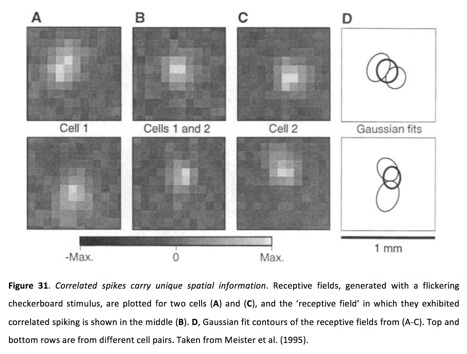
Despite its prevalence, it remains unclear what role correlated ganglion cell activity plays in higher visual processing. One hypothesized role is that it could enhance synaptic transmission in retinorecipient cells. In the lateral geniculate nucleus (LGN), retinal inputs with short inter-spike intervals greatly increase synaptic efficacy (Usrey et al., 1998; Carandini et al., 2007). Considering that multiple neighbouring ganglion cells converge onto individual LGN relay neurons (Cleland et al., 1971; Chen and Regehr, 2000; Morgan et al., 2016; Rompani et al., 2017), ganglion cell synchrony appears to be a highly plausible strategy for effectively driving LGN neurons. Another hypothesized role for ganglion cell synchrony is that it enhances bandwidth of the optic nerve (Meister et al., 1995; Schnitzer and Meister, 2003). Since correlated ganglion cell spikes tend to occur when light activates overlapping areas between neighbouring ganglion cell receptive fields (Figure 31; Meister et al., 1995; Schnitzer and Meister, 2003; Trenholm et al., 2014), correlated activity contains unique spatial information which could subsequently be decoded separately from uncorrelated activity.
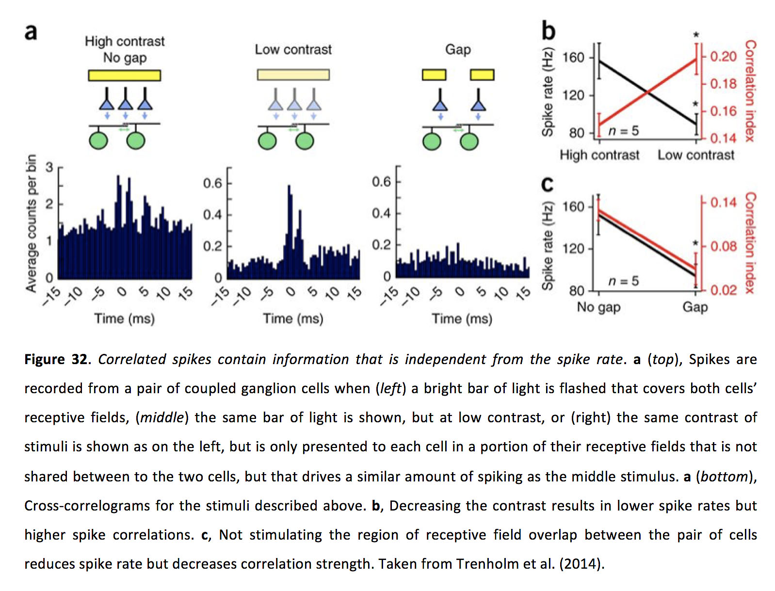
Furthermore, since the strength of narrow gap junction mediated ganglion cell correlations appears to decrease with increasing spike rates (Xiao et al., 2013; Trenholm et al., 2014; though an inhibitory mechanism may sometimes concomitantly lower spike rates and reduce medium scale gap junction mediated synchrony (Ackert et al., 2006)), correlated ganglion cell activity could relay information to downstream targets independent from spike rate (Figure 32). Whether these types of parallel decoding strategies are actually implemented in higher visual areas remains to be examined.
Aside from the above described forms of synchrony, which tend to be restricted to neighbouring retinal neurons, gap junctions also appear to play in an important role in synchronizing activity across wide regions of the retina. Classic work with paired recordings revealed the presence of long-range correlated activity in the retina across distances larger than 20 degrees of visual space (Neuenschwander and Singer, 1996). Such synchrony appeared to arise when pairs of distally located ganglion cells were responding to a visual image that fell upon their receptive fields as well as the space in between them (Neuenschwander and Singer, 1996). More recent work has shown that this long-range synchrony arises from gap junction connectivity between ganglion cells, likely via an intermediary widefield amacrine cell (Roy et al., 2017). Remarkably, as was originally hypothesized (Neuenschwander and Singer, 1996), this long-range synchrony appears to play an important behavioral role in an animal’s ability to bind together distal portions of the same visual image (Roy et al., 2017).
10. Gap junctions and moving visual stimuli
The classical view of the excitatory retinal signaling pathway is of a vertical circuit from photoreceptors to bipolar cells to ganglion cells. Placing gap junctions between neighbouring cells adds a lateral excitatory connection to this vertical pathway. Unsurprisingly, adding such a lateral excitatory pathway endows retinal circuits with distinct advantages when it comes to processing moving visual stimuli.
11. Motion sensitivity
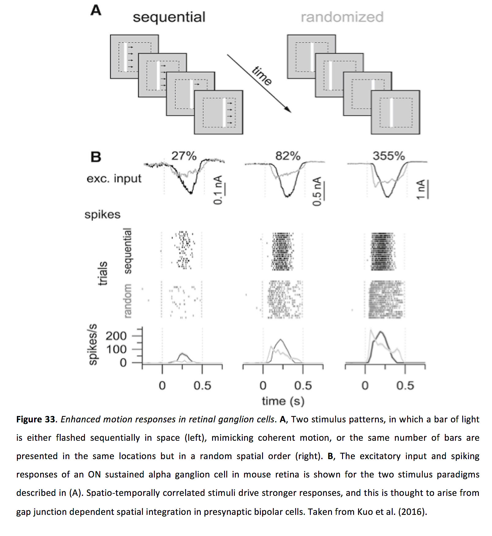
Lateral gap junction connectivity, in both bipolar cells and ganglion cells, has been shown to selectively modify and enhance responses to moving stimuli. In mouse retina, it has been shown that gap junction inputs to bipolar cells (which can represent > 30% of their excitatory synaptic input), can endow bipolar cells with supralinear spatial summation, which selectively enhances processing of spatio-temporally correlated inputs (i.e. a stimuli moving in a coherent direction). This results in enhanced responses to motion in retinal ganglion cells (Figure 33; Kuo et al., 2016). Related work has been done in primate retina, in which bipolar cell gap junction coupling appears to contribute to motion sensitivity of parasol ganglion cells (Manookin et al., 2018). The general principles underlying motion facilitation in gap junction networks in bipolar cells appears to be similar to that described in more detail in a later section on ganglion cells (Murphy-Baum and Awatramani, 2018).
Finally, in mouse retina, at low light levels, homologously coupled direction selective ganglion cells have broader tuning curves than their uncoupled counterparts. As such, gap junction connectivity appears to allow these cells to strike a balance, in low light levels, between detecting dim moving stimuli and accurately reporting the direction of movement (Figure 34; Yao et al., 2018).

12. Motion anticipation
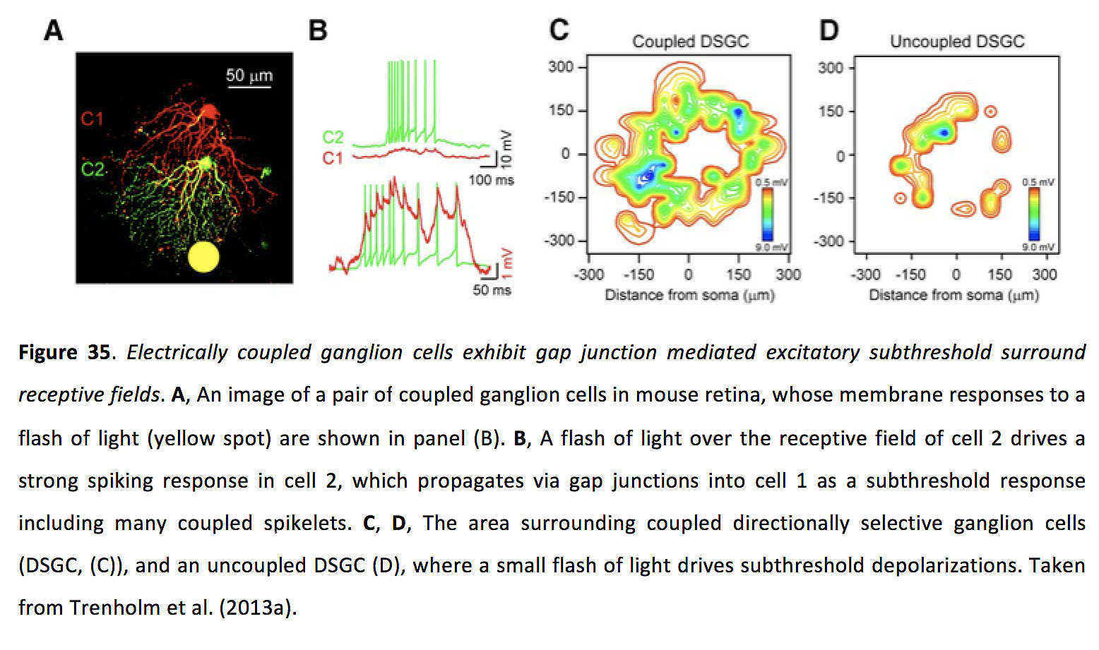
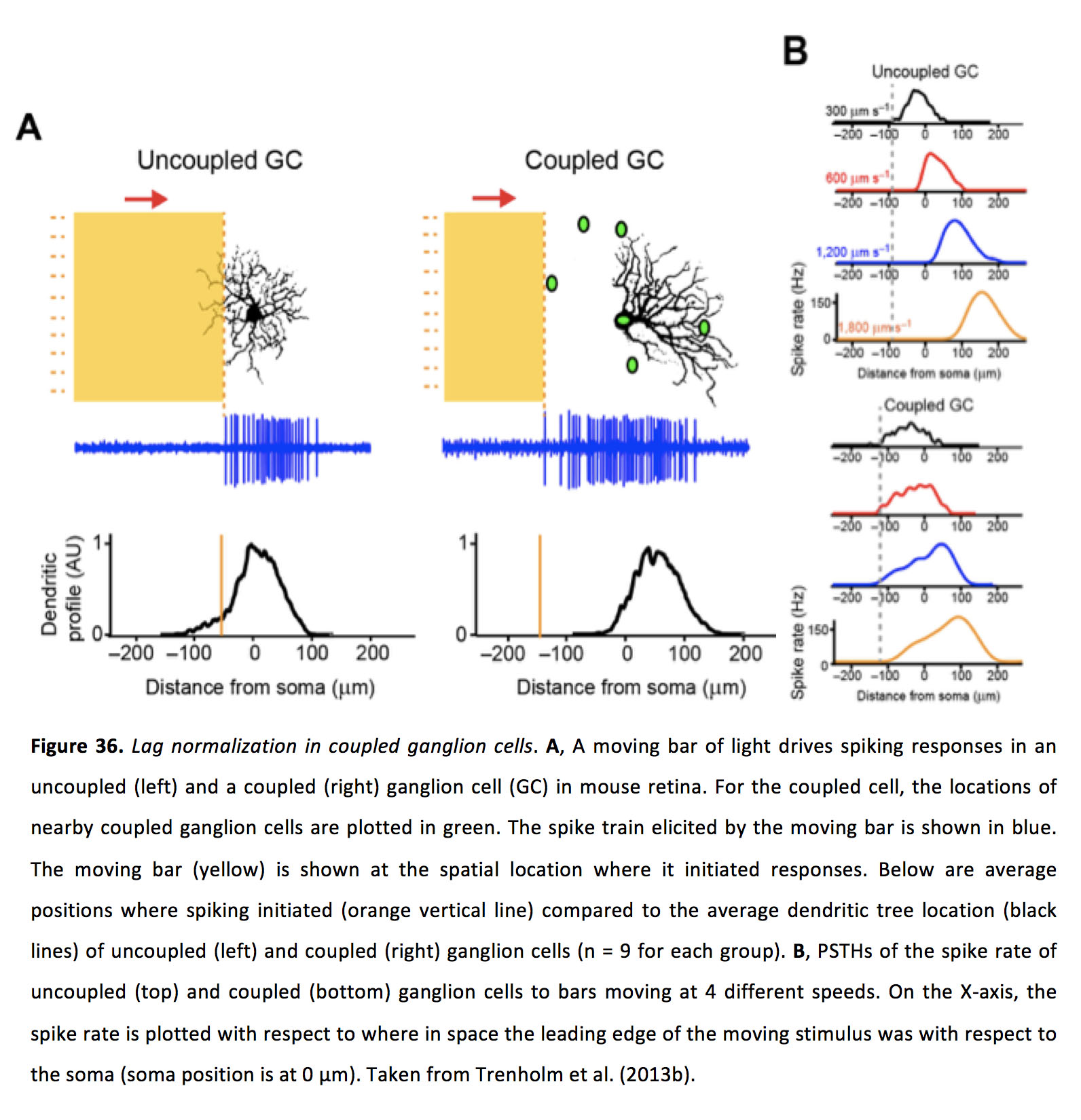
When light is flashed on the retina, it takes roughly 50 ms or more for a ganglion cell to fire a light-evoked action potential. The majority of this delay is due to phototransduction in rods and cones, the relatively slow process whereby incoming photons are converted into an electro-chemical signal. This delay poses a problem, given that the same retinal circuity can process both static and moving stimuli. For static images, this delay does not pose a significant problem, as when a ganglion cell spikes the stimulus it is encoding is still in the appropriate position. In contrast, when a moving image activates photoreceptors in one location, the image will have moved to a different spatial location by the time the ganglion cells vertically connected to the photoreceptors at the earlier position begin to respond. The lag between where a ganglion cell reports an image is compared to where the image actually is in space increases as movement gets faster. One method that has been hypothesized for the retina to address this problem is for ganglion cells to dynamically shift the peak of their receptive field toward the leading edge of a moving stimulus (Berry et al., 1999; Johnston and Lagnado, 2015), which could theoretically be read out by downstream targets as anticipation if they decode position based on peak spike rate. Another method that has been proposed to compensate for the lag that arises when coding moving stimuli involves using gap junctions to allow upstream retinal ganglion cells to alert their downstream neighbors to the presence of an incoming image. This phenomenon, termed lag normalization, arises in homologously coupled retinal ganglion cells which exhibit a coupling-mediated subthreshold excitatory receptive field that surrounds their receptive field center (Figure 35; Trenholm et al., 2013a).
For moving images, the gap junction inputs spread laterally from one ganglion cell to another, and are able to combine with weak bipolar cell mediated chemical synaptic inputs to allow the ganglion cell to begin responding outside of the receptive area that drove spiking activity to static stimuli (Trenholm et al., 2013b). Moreover, when these coupled ganglion cells are stimulated with images moving at various speeds, they can initiate their responses at nearly the same spatial location (i.e. they ‘lag normalize’), in contrast to uncoupled ganglion cells which initiate their responses at positions further and further in space when presented with faster and faster speeds of moving images (Figure 36; Trenholm et al., 2013b).
13. Looming
Effective detection of looming visual stimuli, like an approaching predator, can be a matter of life or death. Accordingly, looming (or approaching) visual stimuli drive behavioral reflexes that are found across many species, including humans (Ball and Tronick, 1971; Schiff et al., 1962; King et al., 1992; Ishikane et al., 2005; Temizer et al., 2015). In the retina, it appears that lateral gap junction connectivity – between AII amacrine cells and ON cone bipolar cells – plays a role in allowing a specific retinal ganglion cell to preferentially detect looming visual images. In mouse retina, the looming sensitive cell is an OFF ganglion cell, termed the PV-5 cell (Münch et al., 2009). When static decrements of light are presented over the receptive field of this cell, they evoke transient OFF excitation, followed by transient ON inhibition when the light level returns to baseline (Münch et al., 2009). As a large dark stimulus gets smaller over the receptive field of this cell, it exclusively drives ON inhibition and no excitatory OFF response, and thus does not result in spiking. On the other hand, if the same stimulus is now played backwards and the black area expands over the receptive field of the cell, it exclusively drives OFF excitation that drives robust spiking responses. Based on the described response properties, this cell should also respond reasonably well to moving stimuli that pass across the receptive field, which would drive a mix of OFF excitation and ON inhibition. However, the ON inhibitory input to the PV-5 cell is very fast, and appears to arise via gap junction signals passed from ON cone bipolar cells to AII amacrine cells, which weaken the effect of concomitant excitatory OFF inputs. Indeed, when Cx36 was knocked out, PV-5 cells no longer responded preferentially to looming stimuli over stimuli that simply moved across the receptive field (Münch et al., 2009). In mice, this retinal cell type is thought to underlie innate freezing/fleeing behavioral responses when dark looming stimuli are presented above the head of a mouse (Yilmaz and Meister, 2013; Yilmaz et al., 2014). Whether similar retinal circuitry underlies behavioral responses to looming visual stimuli in primates remains to be tested.
14. Gap junctions, retinal degeneration and spontaneous activity
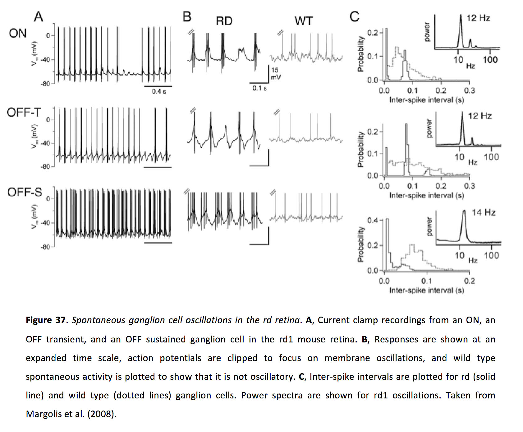
While, as outlined above, gap junctions serve many functions in the healthy retina, they also promote aberrant neuronal activity following retinal degeneration (rd). In rodent rd models, following photoreceptor degeneration, instead of becoming silent many ganglion cells exhibit spontaneous oscillations with a frequency of around 10 Hz (Figure 37; Margolis et al., 2008; Stasheff, 2008; Borowska et al., 2011; Menzler and Zeck, 2011; Stasheff et al., 2011; Yee et al., 2012; Biswas et al., 2014). Understanding the source of this aberrant activity is important since it could underlie phosphenes experienced by patients suffering from vision loss (Lepore, 1990; Murtha and Stasheff, 2003), and could impact the success of therapeutic strategies including electrical implants (Weiland and Humayun, 2014) and optogenetic vision therapy (Busskamp et al., 2012).
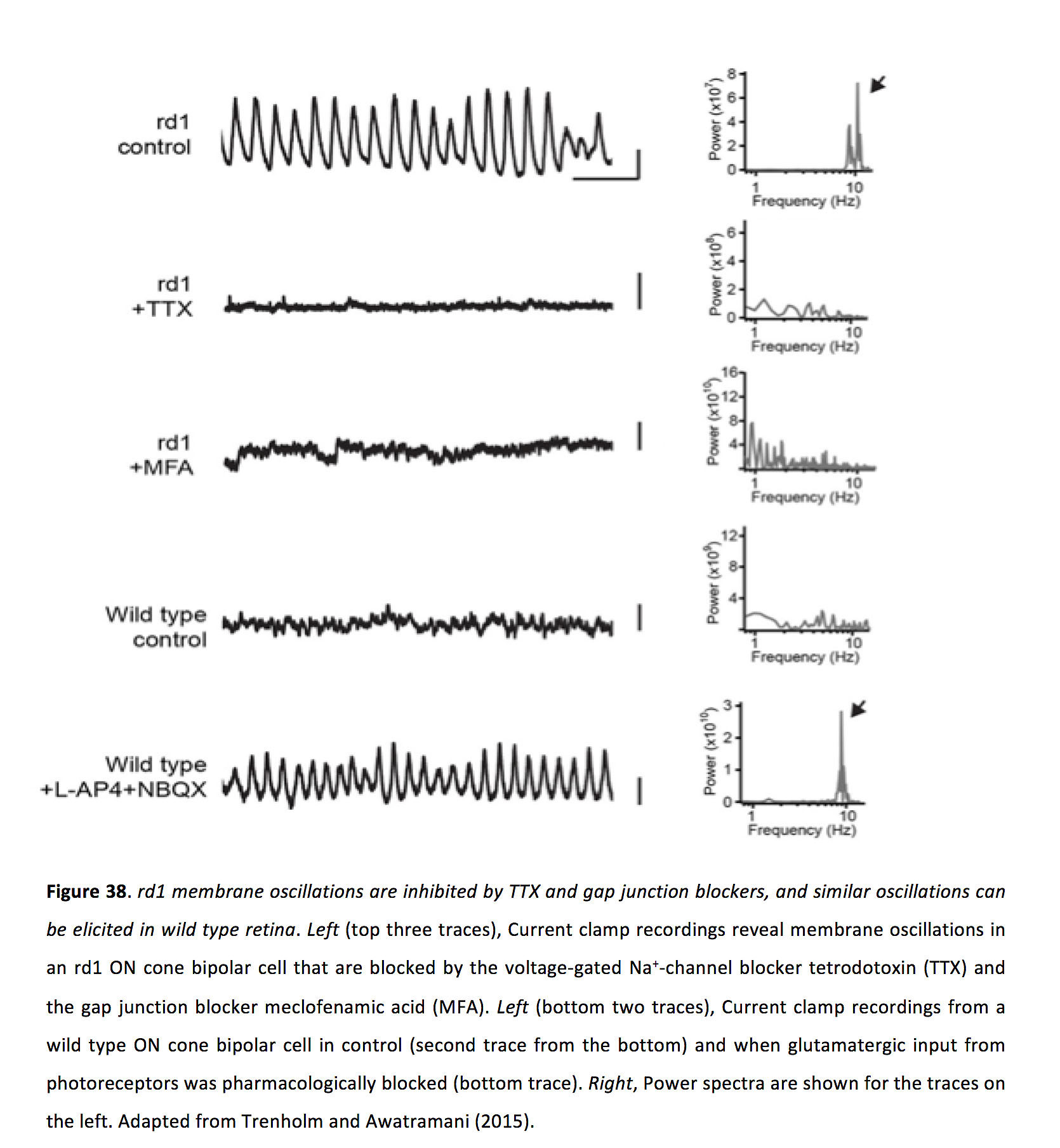
In rd retina, ganglion cells oscillate as the result of receiving excitatory and inhibitory oscillatory inputs (Margolis et al., 2008; Borowska et al., 2011; Yee et al., 2012, 2014). Application of chemical synaptic blockers inhibits ganglion cell oscillations, indicating that spontaneous oscillations do not arise in ganglion cells themselves (Borowska et al., 2011; Menzler and Zeck, 2011; but see also Sekirnjak et al., 2011). In contrast, membrane oscillations in ON cone bipolar cells and AII amacrine cells, which also occur with a frequency of around 10 Hz, persist upon application of chemical synaptic blockers, indicating that spontaneous oscillations arise intrinsically within the coupled network of ON cone bipolar cells and AII amacrine cells (Borowska et al., 2011). As evidence that gap junctions between bipolar cells and AII amacrine cells are important in this pathophysiology, pharmacologically blocking gap junctions inhibits oscillations in ganglion cells (Menzler and Zeck, 2011; Trenholm et al., 2012), AII amacrine cells (Trenholm et al., 2012) and ON cone bipolar cells (Figure 38; Trenholm et al., 2012). Furthermore, spontaneous ganglion cell oscillations were greatly reduced in rd mice in which connexin 36 was knocked out (Ivanova et al., 2016).
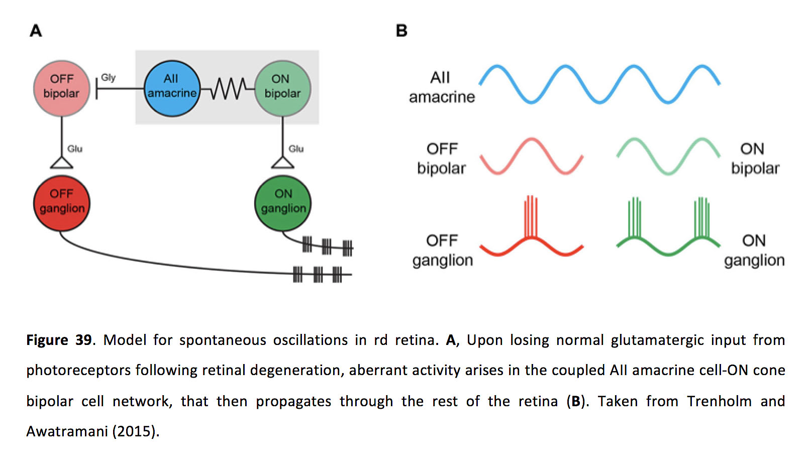
Considering that there is extensive coupling between ON cone bipolar cells and AII amacrine cells in wild type retina, what occurs during retinal degeneration that promotes aberrant network activity? Experiments in wild type retina suggest that the main trigger for spontaneous activity relates to altered glutamate release from photoreceptors, as either pharmacologically blocking photoreceptor output in wild type retina or photo-bleaching the wild type retina with bright light (both of which would hyperpolarize the AII amacrine/ON cone bipolar cell network) can drive spontaneous activity similar to that found in the rd retina (Trenholm et al., 2012; Menzler et al., 2014). Consistent with this idea, the resting membrane potential of AII amacrine cells in the degenerating retina appears to be hyperpolarized compared to those in the wild type retina (Choi et al., 2014). Such hyperpolarization could activate rhythmogenic conductances, such as Ih which is present in some ON cone bipolar cells and has been shown to modulate oscillations in the rd retina (Trenholm et al., 2012). These oscillations have also been shown to be reliant on TTX-sensitive Na+ channels in AII amacrine cells (Trenholm et al., 2012). Finally, it has been shown that gap junctional coupling of AII amacrine cells in the rd retina appears to be increased (as measured via an increase in phosphorylated Cx36), which could exacerbate aberrant activity following photoreceptor degeneration (Ivanova et al., 2015). As such, membrane conductances and gap junctions in the AII amacrine and ON bipolar cell network act in tandem following photoreceptor degeneration to elicit spontaneous membrane oscillations (Figure 39).
15. Conclusions
It is well established that the retina is extensively gap junction coupled, and that these connections endow retinal circuits with a plethora of functional properties. In the retina, gap junctions are associated with night vision, controlling receptive field size, signal correlation, spatial integration, and motion detection. Despite these findings, many unknowns remain. First, many critical experiments into the roles of gap junctions in the retina have employed either pharmacological agents or Cx36 KO to examine the roles of gap junctions in retinal circuits. However, simultaneously blocking gap junctions at all levels of the retina makes it difficult to assign roles for specific electrical synapses between given cell types. As such, future research will need to endeavor to modify gap junctions in specific cell types (eg. Yao et al., 2018). Second, related to the first point, while it is clear that the retina splits incoming visual signals into approximately 30 distinct retinal channels (made up of distinct cell types, connected in distinct ways), unique roles for gap junctions in each of these channels is only beginning to be studied. Third, given that gap junctions are known to play important roles in retinal development (Cook and Becker, 2009), and may affect susceptibility to disease pathologies (Striedinger et al., 2005; Paschon et al., 2012; Kranz et al., 2013; Akopian et al., 2014), it will be important to examine whether gap junctions play precise cell-type-specific roles in development (Arroyo et al., 2016) and disease. Finally, in addition to increasing our knowledge into the fine details of roles for gap junctions in microcircuits, it will be important to characterize different manners in which gap junction connections are plastic, and thus allow circadian rhythm, light adaptation, or neuronal activity to gate their activity and modify circuit function.
Acknowledgements
This work was supported by Canada Research Chairs to ST and GBA.
About the authors

Dr. Stuart Trenholm received his PhD in neuroscience from Dalhousie University, in Halifax Canada, in the lab of Dr. Gautam Awatramani. Subsequently he was a postdoctoral fellow in neuroscience in the lab of Dr. Botond Roska, at the Friedrich Miescher Institute for Biomedical Research in Basel, Switzerland. He is now an Assistant Professor at the Montreal Neurological Institute at McGill University, in Montreal, Canada. His lab examines the neuronal circuitry underlying visual perception and studies vision rehabilitation strategies.

Dr. Gautam Awatramani received his B.S. from the University of Rochester (1995) and then did his Ph.D. in Physiology and Biophysics under the supervision of Dr. Malcolm Slaughter at SUNY Buffalo. He was a postdoctoral fellow at the Vollum Institute (Portland, OR, 2000-2004) with Dr. Laurence Trussell, at the University of British Columbia (Vancouver, B.C, 2005-2007) with Dr. Tim Murphy and at the Friedrich Miescher Institute (Basel, CH, 2008) with Dr. Botond Roska. He then started his own lab at Dalhousie University (Halifax 2008-2011) and subsequently moved to the University of Victoria (Victoria, B.C.) where he is currently an Associate Professor/CRC chair in Physiology. His work currently focusses on understanding the synaptic mechanisms underlying the generation of direction selectivity in the retina.
References
1. Masland R.H. The fundamental plan of the retina. Nature Neuroscience. 2001;4(9):877–886. [PubMed]
2. Gollisch T., Meister M. Eye smarter than scientists believed: neural computations in circuits of the retina. Neuron. 2010;65(2):150–164. [PMC free article] [PubMed]
3. Azeredo da Silveira R., Roska B. Cell types, circuits, computation. Current Opinion in Neurobiology. 2011;21(5):664–671. [PubMed]
4. Masland R.H. The neuronal organization of the retina. Neuron. 2012;76(2):266–280. [PMC free article] [PubMed]
5. Diamond J.S. Inhibitory Interneurons in the Retina: Types, Circuitry, and Function. Annual Review of Vision Science. 2017;3:1–24. [PubMed]
6. Martersteck E.M., Hirokawa K.E., Evarts M., Bernard A., Duan X., Li Y., Ng L., Oh S.W., Ouellette B., Royall J.J., Stoecklin M., Wang Q., Zeng H., Sanes J.R., Harris J.A. Diverse Central Projection Patterns of Retinal Ganglion Cells. Cell Reports. 2017;18(8):2058–2072. [PMC free article] [PubMed]
7. Robles E., Laurell E., Baier H. The retinal projectome reveals brain-area-specific visual representations generated by ganglion cell diversity. Current biology: CB. 2014;24(18):2085–2096. [PubMed]
8. Ribelayga, C.P. and J. O’Brien, Chapter 10 – Circadian and Light-Adaptive Control of Electrical Synaptic Plasticity in the Vertebrate Retina, in Network Functions and Plasticity, J. Jing, Editor. 2017, Academic Press. p. 209-241.
9. Baylor D.A., Fuortes M.G., O’Bryan P.M. Receptive fields of cones in the retina of the turtle. The Journal of Physiology. 1971;214(2):265–294. [PMC free article] [PubMed]
10. Copenhagen D.R., Owen W.G. Coupling between rod photoreceptors in a vertebrate retina. Nature. 1976;260(5546):57–59. [PubMed]
11. Custer N.V. Structurally specialized contacts between the photoreceptors of the retina of the axolotl. The Journal of Comparative Neurology. 1973;151(1):35–56. [PubMed]
12. DeVries S.H., Qi X., Smith R., Makous W., Sterling P. Electrical coupling between mammalian cones. Current biology: CB. 2002;12(22):1900–1907. [PubMed]
13. Hornstein E.P., Verweij J., Li P.H., Schnapf J.L. Gap-junctional coupling and absolute sensitivity of photoreceptors in macaque retina. The Journal of Neuroscience: The Official Journal of the Society for Neuroscience. 2005;25(48):11201–11209. [PMC free article] [PubMed]
14. Hornstein E.P., Verweij J., Schnapf J.L. Electrical coupling between red and green cones in primate retina. Nature Neuroscience. 2004;7(7):745–750. [PubMed]
15. Li P.H., Verweij J., Long J.H., Schnapf J.L. Gap-junctional coupling of mammalian rod photoreceptors and its effect on visual detection. The Journal of Neuroscience: The Official Journal of the Society for Neuroscience. 2012;32(10):3552–3562. [PMC free article] [PubMed]
16. Raviola E., Gilula N.B. Gap junctions between photoreceptor cells in the vertebrate retina. Proceedings of the National Academy of Sciences of the United States of America. 1973;70(6):1677–1681. [PMC free article] [PubMed]
17. Tsukamoto Y., Morigiwa K., Ueda M., Sterling P. Microcircuits for night vision in mouse retina. The Journal of Neuroscience: The Official Journal of the Society for Neuroscience. 2001;21(21):8616–8623. [PMC free article] [PubMed]
18. Feigenspan A., Janssen-Bienhold U., Hormuzdi S., Monyer H., Degen J., Söhl G., Willecke K., Ammermüller J., Weiler R. Expression of connexin36 in cone pedicles and OFF-cone bipolar cells of the mouse retina. The Journal of Neuroscience: The Official Journal of the Society for Neuroscience. 2004;24(13):3325–3334. [PMC free article] [PubMed]
19. Kántor O., Benkő Z., Énzsöly A., Dávid C., Naumann A., Nitschke R., Szabó A., Pálfi E., Orbán J., Nyitrai M., Németh J., Szél Á., Lukáts Á., Völgyi B. Characterization of connexin36 gap junctions in the human outer retina. Brain Structure & Function. 2016;221(6):2963–2984. [PubMed]
20. Lee E.-J., Han J.-W., Kim H.-J., Kim I.-B., Lee M.-Y., Oh S.-J., Chung J.-W., Chun M.-H. The immunocytochemical localization of connexin 36 at rod and cone gap junctions in the guinea pig retina. The European Journal of Neuroscience. 2003;18(11):2925–2934. [PubMed]
21. O’Brien J.J., Chen X., Macleish P.R., O’Brien J., Massey S.C. Photoreceptor coupling mediated by connexin36 in the primate retina. The Journal of Neuroscience: The Official Journal of the Society for Neuroscience. 2012;32(13):4675–4687. [PMC free article] [PubMed]
22. Li H., Chuang A.Z., O’Brien J. Photoreceptor coupling is controlled by connexin 35 phosphorylation in zebrafish retina. The Journal of Neuroscience: The Official Journal of the Society for Neuroscience. 2009;29(48):15178–15186. [PMC free article] [PubMed]
23. Li H., Zhang Z., Blackburn M.R., Wang S.W., Ribelayga C.P., O’Brien J. Adenosine and dopamine receptors coregulate photoreceptor coupling via gap junction phosphorylation in mouse retina. The Journal of Neuroscience: The Official Journal of the Society for Neuroscience. 2013;33(7):3135–3150. [PMC free article] [PubMed]
24. Zhang J., Wu S.M. Connexin35/36 gap junction proteins are expressed in photoreceptors of the tiger salamander retina. The Journal of Comparative Neurology. 2004;470(1):1–12. [PubMed]
25. Asteriti S., Gargini C., Cangiano L. Connexin 36 expression is required for electrical coupling between mouse rods and cones. Visual Neuroscience. 2017;34:E006 [PubMed]
26. Kolb H. The organization of the outer plexiform layer in the retina of the cat: electron microscopic observations. Journal of Neurocytology. 1977;6(2):131–153. [PubMed]
27. Li W., DeVries S.H. Separate blue and green cone networks in the mammalian retina. Nature Neuroscience. 2004;7(7):751–756. [PubMed]
28. Tsukamoto Y., Masarachia P., Schein S.J., Sterling P. Gap junctions between the pedicles of macaque foveal cones. Vision Research. 1992;32(10):1809–1815. [PubMed]
29. Zhang J., Wu S.M. Physiological properties of rod photoreceptor electrical coupling in the tiger salamander retina. The Journal of Physiology. 2005;564(Pt 3):849–862. [PMC free article] [PubMed]
30. Werblin F.S. Transmission along and between rods in the tiger salamander retina. The Journal of Physiology. 1978;280:449–470. [PMC free article] [PubMed]
31. Gao F., Pang J.-J., Wu S.M. Sign-preserving and sign-inverting synaptic interactions between rod and cone photoreceptors in the dark-adapted retina. The Journal of Physiology. 2013;591(22):5711–5726. [PMC free article] [PubMed]
32. Attwell D., Wilson M., Wu S.M. A quantitative analysis of interactions between photoreceptors in the salamander (Ambystoma) retina. The Journal of Physiology. 1984;352:703–737. [PMC free article] [PubMed]
33. Ribelayga C., Cao Y., Mangel S.C. The circadian clock in the retina controls rod-cone coupling. Neuron. 2008;59(5):790–801. [PMC free article] [PubMed]
34. Wu S.M., Yang X.L. Electrical coupling between rods and cones in the tiger salamander retina. Proceedings of the National Academy of Sciences of the United States of America. 1988;85(1):275–278. [PMC free article] [PubMed]
35. Asteriti S., Gargini C., Cangiano L. Mouse rods signal through gap junctions with cones. eLife. 2014;3:e01386 [PMC free article] [PubMed]
36. Nelson R. Cat cones have rod input: a comparison of the response properties of cones and horizontal cell bodies in the retina of the cat. The Journal of Comparative Neurology. 1977;172(1):109–135. [PubMed]
37. Schneeweis D.M., Schnapf J.L. Photovoltage of rods and cones in the macaque retina. Science (New York, N.Y.). 1995;268(5213):1053–1056. [PubMed]
38. Thoreson W.B., Mangel S.C. Lateral interactions in the outer retina. Progress in Retinal and Eye Research. 2012;31(5):407–441. [PMC free article] [PubMed]
39. Raviola E., Gilula N.B. Intramembrane organization of specialized contacts in the outer plexiform layer of the retina. A freeze-fracture study in monkeys and rabbits. The Journal of Cell Biology. 1975;65(1):192–222. [PMC free article] [PubMed]
40. Hombach S., Janssen-Bienhold U., Söhl G., Schubert T., Büssow H., Ott T., Weiler R., Willecke K. Functional expression of connexin57 in horizontal cells of the mouse retina. The European Journal of Neuroscience. 2004;19(10):2633–2640. [PubMed]
41. Janssen-Bienhold U., Trümpler J., Hilgen G., Schultz K., Müller L.P.D.S., Sonntag S., Dedek K., Dirks P., Willecke K., Weiler R. Connexin57 is expressed in dendro-dendritic and axo-axonal gap junctions of mouse horizontal cells and its distribution is modulated by light. The Journal of Comparative Neurology. 2009;513(4):363–374. [PubMed]
42. Dorgau B., Herrling R., Schultz K., Greb H., Segelken J., Ströh S., Bolte P., Weiler R., Dedek K., Janssen-Bienhold U. Connexin50 couples axon terminals of mouse horizontal cells by homotypic gap junctions. The Journal of Comparative Neurology. 2015;523(14):2062–2081. [PubMed]
43. O’Brien J.J., Li W., Pan F., Keung J., O’Brien J., Massey S.C. Coupling between A-type horizontal cells is mediated by connexin 50 gap junctions in the rabbit retina. The Journal of Neuroscience: The Official Journal of the Society for Neuroscience. 2006;26(45):11624–11636. [PMC free article] [PubMed]
44. Pan F., Keung J., Kim I.-B., Snuggs M.B., Mills S.L., O’Brien J., Massey S.C. Connexin 57 is expressed by the axon terminal network of B-type horizontal cells in the rabbit retina. The Journal of Comparative Neurology. 2012;520(10):2256–2274. [PMC free article] [PubMed]
45. Klaassen L.J., Sun Z., Steijaert M.N., Bolte P., Fahrenfort I., Sjoerdsma T., Klooster J., Claassen Y., Shields C.R., Ten Eikelder H.M.M., Janssen-Bienhold U., Zoidl G., McMahon D.G., Kamermans M. Synaptic transmission from horizontal cells to cones is impaired by loss of connexin hemichannels. PLoS biology. 2011;9(7):e1001107 [PMC free article] [PubMed]
46. Shields C.R., Klooster J., Claassen Y., Ul-Hussain M., Zoidl G., Dermietzel R., Kamermans M. Retinal horizontal cell-specific promoter activity and protein expression of zebrafish connexin 52.6 and connexin 55.5. The Journal of Comparative Neurology. 2007;501(5):765–779. [PubMed]
47. DeVries S.H., Schwartz E.A. Modulation of an electrical synapse between solitary pairs of catfish horizontal cells by dopamine and second messengers. The Journal of Physiology. 1989;414:351–375. [PMC free article] [PubMed]
48. Lasater E.M., Dowling J.E. Dopamine decreases conductance of the electrical junctions between cultured retinal horizontal cells. Proceedings of the National Academy of Sciences of the United States of America. 1985;82(9):3025–3029. [PMC free article] [PubMed] 49. McMahon D.G. Modulation of electrical synaptic transmission in zebrafish retinal horizontal cells. The Journal of Neuroscience: The Official Journal of the Society for Neuroscience. 1994;14(3 Pt 2):1722–1734. [PMC free article] [PubMed] 50. McMahon D.G., Mattson M.P. Horizontal cell electrical coupling in the giant danio: synaptic modulation by dopamine and synaptic maintenance by calcium. Brain Research. 1996;718(1-2):89–96. [PubMed] 51. Xin D., Bloomfield S.A. Dark- and light-induced changes in coupling between horizontal cells in mammalian retina. The Journal of Comparative Neurology. 1999;405(1):75–87. [PubMed]
52. Ribelayga C., Mangel S.C. Absence of circadian clock regulation of horizontal cell gap junctional coupling reveals two dopamine systems in the goldfish retina. The Journal of Comparative Neurology. 2003;467(2):243–253. [PubMed]
53. Ribelayga C., Mangel S.C. Tracer coupling between fish rod horizontal cells: modulation by light and dopamine but not the retinal circadian clock. Visual Neuroscience. 2007;24(3):333–344. [PubMed]
54. Mangel S.C., Dowling J.E. Responsiveness and receptive field size of carp horizontal cells are reduced by prolonged darkness and dopamine. Science (New York, N.Y.). 1985;229(4718):1107–1109. [PubMed]
55. Shelley J., Dedek K., Schubert T., Feigenspan A., Schultz K., Hombach S., Willecke K., Weiler R. Horizontal cell receptive fields are reduced in connexin57-deficient mice. The European Journal of Neuroscience. 2006;23(12):3176–3186. [PubMed]
56. Mangel S.C. Analysis of the horizontal cell contribution to the receptive field surround of ganglion cells in the rabbit retina. The Journal of physiology. 1991;442(1):211–234. [PMC free article] [PubMed]
57. Werblin F.S., Dowling J.E. Organization of the retina of the mudpuppy, Necturus maculosus. II. Intracellular recording. Journal of Neurophysiology. 1969;32(3):339–355. [PubMed]
58. Drinnenberg A., Franke F., Morikawa R.K., Jüttner J., Hillier D., Hantz P., Hierlemann A., Azeredo da Silveira R., Roska B. How Diverse Retinal Functions Arise from Feedback at the First Visual Synapse. Neuron. 2018;99(1):117–134.e11. [PMC free article] [PubMed]
59. Farrow K., Teixeira M., Szikra T., Viney T.J., Balint K., Yonehara K., Roska B. Ambient illumination toggles a neuronal circuit switch in the retina and visual perception at cone threshold. Neuron. 2013;78(2):325–338. [PubMed]
60. Hoggarth A., McLaughlin A.J., Ronellenfitch K., Trenholm S., Vasandani R., Sethuramanujam S., Schwab D., Briggman K.L., Awatramani G.B. Specific wiring of distinct amacrine cells in the directionally selective retinal circuit permits independent coding of direction and size. Neuron. 2015;86(1):276–291. [PubMed]
61. Dedek K., Pandarinath C., Alam N.M., Wellershaus K., Schubert T., Willecke K., Prusky G.T., Weiler R., Nirenberg S. Ganglion cell adaptability: does the coupling of horizontal cells play a role? PloS One. 2008;3(3):e1714 [PMC free article] [PubMed]
62. Pandarinath C., Bomash I., Victor J.D., Prusky G.T., Tschetter W.W., Nirenberg S. A novel mechanism for switching a neural system from one state to another. Frontiers in Computational Neuroscience. 2010;4:2. [PMC free article] [PubMed]
63. Wässle H., Puller C., Müller F., Haverkamp S. Cone contacts, mosaics, and territories of bipolar cells in the mouse retina. The Journal of Neuroscience: The Official Journal of the Society for Neuroscience. 2009;29(1):106–117. [PMC free article] [PubMed]
64. Kolb H., Famiglietti E.V. Rod and cone pathways in the inner plexiform layer of cat retina. Science (New York, N.Y.). 1974;186(4158):47–49. [PubMed]
65. Lee S.C.S., Meyer A., Schubert T., Hüser L., Dedek K., Haverkamp S. Morphology and connectivity of the small bistratified A8 amacrine cell in the mouse retina. The Journal of Comparative Neurology. 2015;523(10):1529–1547. [PMC free article] [PubMed]
66. Arai I., Tanaka M., Tachibana M. Active roles of electrically coupled bipolar cell network in the adult retina. The Journal of Neuroscience: The Official Journal of the Society for Neuroscience. 2010;30(27):9260–9270. [PMC free article] [PubMed]
67. Jacoby R.A., Marshak D.W. Synaptic connections of DB3 diffuse bipolar cell axons in macaque retina. The Journal of Comparative Neurology. 2000;416(1):19–29. [PMC free article] [PubMed]
68. Marc R.E., Liu W.L., Muller J.F. Gap junctions in the inner plexiform layer of the goldfish retina. Vision Research. 1988;28(1):9–24. [PubMed]
69. Strettoi E., Raviola E., Dacheux R.F. Synaptic connections of the narrow-field, bistratified rod amacrine cell (AII) in the rabbit retina. The Journal of Comparative Neurology. 1992;325(2):152–168. [PubMed]
70. Deans M.R., Volgyi B., Goodenough D.A., Bloomfield S.A., Paul D.L. Connexin36 is essential for transmission of rod-mediated visual signals in the mammalian retina. Neuron. 2002;36(4):703–712. [PMC free article] [PubMed]
71. Han Y., Massey S.C. Electrical synapses in retinal ON cone bipolar cells: subtype-specific expression of connexins. Proceedings of the National Academy of Sciences of the United States of America. 2005;102(37):13313–13318. [PMC free article] [PubMed]
72. Hilgen G., von Maltzahn J., Willecke K., Weiler R., Dedek K. Subcellular distribution of connexin45 in OFF bipolar cells of the mouse retina. The Journal of Comparative Neurology. 2011;519(3):433–450. [PubMed]
73. Maxeiner S., Dedek K., Janssen-Bienhold U., Ammermüller J., Brune H., Kirsch T., Pieper M., Degen J., Krüger O., Willecke K., Weiler R. Deletion of connexin45 in mouse retinal neurons disrupts the rod/cone signaling pathway between AII amacrine and ON cone bipolar cells and leads to impaired visual transmission. The Journal of Neuroscience: The Official Journal of the Society for Neuroscience. 2005;25(3):566–576. [PMC free article] [PubMed]
74. Dacey D., Packer O.S., Diller L., Brainard D., Peterson B., Lee B. Center surround receptive field structure of cone bipolar cells in primate retina. Vision Research. 2000;40(14):1801–1811. [PubMed]
75. Kujiraoka T., Saito T. Electrical coupling between bipolar cells in carp retina. Proceedings of the National Academy of Sciences of the United States of America. 1986;83(11):4063–4066. [PMC free article] [PubMed]
76. Mills S.L. Unusual coupling patterns of a cone bipolar cell in the rabbit retina. Visual Neuroscience. 1999;16(6):1029–1035. [PubMed]
77. Umino O., Maehara M., Hidaka S., Kita S., Hashimoto Y. The network properties of bipolar-bipolar cell coupling in the retina of teleost fishes. Visual Neuroscience. 1994;11(3):533–548. [PubMed]
78. Veruki M.L., Hartveit E. AII (Rod) amacrine cells form a network of electrically coupled interneurons in the mammalian retina. Neuron. 2002;33(6):935–946. [PubMed]
79. Marc R.E., Anderson J.R., Jones B.W., Sigulinsky C.L., Lauritzen J.S. The AII amacrine cell connectome: a dense network hub. Frontiers in Neural Circuits. 2014;8:104. [PMC free article] [PubMed]
80. Graydon C.W., Lieberman E.E., Rho N., Briggman K.L., Singer J.H., Diamond J.S. Synaptic Transfer between Rod and Cone Pathways Mediated by AII Amacrine Cells in the Mouse Retina. Current biology: CB. 2018;28(17):2739–2751.e3. [PMC free article] [PubMed]
81. Saito T., Kujiraoka T. Characteristics of bipolar-bipolar coupling in the carp retina. The Journal of General Physiology. 1988;91(2):275–287. [PMC free article] [PubMed]
82. Berntson A., Taylor W.R. Response characteristics and receptive field widths of on-bipolar cells in the mouse retina. The Journal of Physiology. 2000;524(Pt 3):879–889. [PMC free article] [PubMed]
83. Werblin F.S. Response of retinal cells to moving spots: intracellular recording in Necturus maculosus. Journal of Neurophysiology. 1970;33(3):342–350. [PubMed]
84. Kuo S.P., Schwartz G.W., Rieke F. Nonlinear Spatiotemporal Integration by Electrical and Chemical Synapses in the Retina. Neuron. 2016;90(2):320–332. [PMC free article] [PubMed]
85. Masland R.H. The tasks of amacrine cells. Visual Neuroscience. 2012;29(1):3–9. [PMC free article] [PubMed]
86. Masland R.H., Mills J.W. Autoradiographic identification of acetylcholine in the rabbit retina. The Journal of Cell Biology. 1979;83(1):159–178. [PMC free article] [PubMed]
87. Lee S., Kim K., Zhou Z.J. Role of ACh-GABA cotransmission in detecting image motion and motion direction. Neuron. 2010;68(6):1159–1172. [PMC free article] [PubMed]
88. Sethuramanujam S., McLaughlin A.J., deRosenroll G., Hoggarth A., Schwab D.J., Awatramani G.B. A Central Role for Mixed Acetylcholine/GABA Transmission in Direction Coding in the Retina. Neuron. 2016;90(6):1243–1256. [PubMed]
89. Zhang D.-Q., Zhou T.-R., McMahon D.G. Functional heterogeneity of retinal dopaminergic neurons underlying their multiple roles in vision. The Journal of Neuroscience: The Official Journal of the Society for Neuroscience. 2007;27(3):692–699. [PMC free article] [PubMed]
90. Baden T., Euler T. Retinal Physiology: Non-Bipolar-Cell Excitatory Drive in the Inner Retina. Current biology: CB. 2016;26(15):R706–R708. [PubMed]
91. Xin D., Bloomfield S.A. Tracer coupling pattern of amacrine and ganglion cells in the rabbit retina. The Journal of Comparative Neurology. 1997;383(4):512–528. [PubMed]
92. Li W., Zhang J., Massey S.C. Coupling pattern of S1 and S2 amacrine cells in the rabbit retina. Visual Neuroscience. 2002;19(2):119–131. [PubMed]
93. Abdel-Majid R.M., Archibald M.L., Tremblay F., Baldridge W.H. Tracer coupling of neurons in the rat retina inner nuclear layer labeled by Fluorogold. Brain Research. 2005;1063(2):114–120. [PubMed]
94. Bloomfield S.A., Xin D. A comparison of receptive-field and tracer-coupling size of amacrine and ganglion cells in the rabbit retina. Visual Neuroscience. 1997;14(6):1153–1165. [PubMed]
95. Völgyi B., Chheda S., Bloomfield S.A. Tracer coupling patterns of the ganglion cell subtypes in the mouse retina. The Journal of Comparative Neurology. 2009;512(5):664–687. [PMC free article] [PubMed]
96. Pan F., Paul D.L., Bloomfield S.A., Völgyi B. Connexin36 is required for gap junctional coupling of most ganglion cell subtypes in the mouse retina. The Journal of Comparative Neurology. 2010;518(6):911–927. [PMC free article] [PubMed]
97. Pang J.-J., Paul D.L., Wu S.M. Survey on amacrine cells coupling to retrograde-identified ganglion cells in the mouse retina. Investigative Ophthalmology & Visual Science. 2013;54(8):5151–5162. [PMC free article] [PubMed]
98. Jacoby J., Nath A., Jessen Z.F., Schwartz G.W. A Self-Regulating Gap Junction Network of Amacrine Cells Controls Nitric Oxide Release in the Retina. Neuron. 2018;100(5):1149–1162.e5. [PMC free article] [PubMed]
99. Grimes W.N., Zhang J., Graydon C.W., Kachar B., Diamond J.S. Retinal parallel processors: more than 100 independent microcircuits operate within a single interneuron. Neuron. 2010;65(6):873–885. [PMC free article] [PubMed]
100. Yadav S.C., Tetenborg S., Dedek K. Gap Junctions in A8 Amacrine Cells Are Made of Connexin36 but Are Differently Regulated Than Gap Junctions in AII Amacrine Cells. Frontiers in Molecular Neuroscience. 2019;12:99. [PMC free article] [PubMed]
101. Dacey, D., Origins of perception: retinal ganglion cell diversity and the creation of parallel visual pathways, in The Cognitive Neurosciences. 2004, MIT Press.
102. Sanes J.R., Masland R.H. The types of retinal ganglion cells: current status and implications for neuronal classification. Annual Review of Neuroscience. 2015;38:221–246. [PubMed]
103. Baden T., Berens P., Franke K., Román Rosón M., Bethge M., Euler T. The functional diversity of retinal ganglion cells in the mouse. Nature. 2016;529(7586):345–350. [PMC free article] [PubMed]
104. Vaney D.I. Many diverse types of retinal neurons show tracer coupling when injected with biocytin or Neurobiotin. Neuroscience Letters. 1991;125(2):187–190. [PubMed]
105. Vaney D.I. Territorial organization of direction-selective ganglion cells in rabbit retina. The Journal of Neuroscience: The Official Journal of the Society for Neuroscience. 1994;14(11 Pt 1):6301–6316. [PMC free article] [PubMed]
106. Hidaka S., Akahori Y., Kurosawa Y. Dendrodendritic electrical synapses between mammalian retinal ganglion cells. The Journal of Neuroscience: The Official Journal of the Society for Neuroscience. 2004;24(46):10553–10567. [PMC free article] [PubMed]
107. Müller L.P.d.S., Do M.T.H., Yau K.-W., He S., Baldridge W.H. Tracer coupling of intrinsically photosensitive retinal ganglion cells to amacrine cells in the mouse retina. The Journal of Comparative Neurology. 2010;518(23):4813–4824. [PMC free article] [PubMed]
108. Reifler A.N., Chervenak A.P., Dolikian M.E., Benenati B.A., Li B.Y., Wachter R.D., Lynch A.M., Demertzis Z.D., Meyers B.S., Abufarha F.S., Jaeckel E.R., Flannery M.P., Wong K.Y. All Spiking, Sustained ON Displaced Amacrine Cells Receive Gap-Junction Input from Melanopsin Ganglion Cells. Current biology: CB. 2015;25(21):2878. [PubMed]
109. Schubert T., Maxeiner S., Krüger O., Willecke K., Weiler R. Connexin45 mediates gap junctional coupling of bistratified ganglion cells in the mouse retina. The Journal of Comparative Neurology. 2005;490(1):29–39. [PubMed]
110. Kántor O., Szarka G., Benkő Z., Somogyvári Z., Pálfi E., Baksa G., Rácz G., Nitschke R., Debertin G., Völgyi B. Strategic Positioning of Connexin36 Gap Junctions Across Human Retinal Ganglion Cell Dendritic Arbors. Frontiers in Cellular Neuroscience. 2018;12:409. [PMC free article] [PubMed]
111. Yao X., Cafaro J., McLaughlin A.J., Postma F.R., Paul D.L., Awatramani G., Field G.D. Gap Junctions Contribute to Differential Light Adaptation across Direction-Selective Retinal Ganglion Cells. Neuron. 2018;100(1):216–228.e6. [PMC free article] [PubMed]
112. Hu E.H., Bloomfield S.A. Gap junctional coupling underlies the short-latency spike synchrony of retinal alpha ganglion cells. The Journal of Neuroscience: The Official Journal of the Society for Neuroscience. 2003;23(17):6768–6777. [PMC free article] [PubMed]
113. Trenholm S., McLaughlin A.J., Schwab D.J., Awatramani G.B. Dynamic tuning of electrical and chemical synaptic transmission in a network of motion coding retinal neurons. The Journal of Neuroscience: The Official Journal of the Society for Neuroscience. 2013;33(37):14927–14938. [PMC free article] [PubMed]
114. Cooler, S. and G.W. Schwartz, An on-primary retinal ganglion cell receives off input via a heterotypic RGC gap junction, in Annual Meeting of the Society for Neuroscience. 2019, Society for Neuroscience: Chicago. p. 225.08.
115. Hu E.H., Pan F., Völgyi B., Bloomfield S.A. Light increases the gap junctional coupling of retinal ganglion cells. The Journal of Physiology. 2010;588(Pt 21):4145–4163. [PMC free article] [PubMed]
116. DeVries S.H. Correlated firing in rabbit retinal ganglion cells. Journal of Neurophysiology. 1999;81(2):908–920. [PubMed]
117. Barlow H.B., Fitzhugh R., Kuffler S.W. Change of organization in the receptive fields of the cat’s retina during dark adaptation. The Journal of Physiology. 1957;137(3):338–354. [PMC free article] [PubMed]
118. Peichl L., Wässle H. The structural correlate of the receptive field centre of alpha ganglion cells in the cat retina. The Journal of Physiology. 1983;341:309–324. [PMC free article] [PubMed]
119. Muller J.F., Dacheux R.F. Alpha ganglion cells of the rabbit retina lose antagonistic surround responses under dark adaptation. Visual Neuroscience. 1997;14(2):395–401. [PubMed]
120. Trong P.K., Rieke F. Origin of correlated activity between parasol retinal ganglion cells. Nature Neuroscience. 2008;11(11):1343–1351. [PMC free article] [PubMed]
121. Nelson R. AII amacrine cells quicken time course of rod signals in the cat retina. Journal of Neurophysiology. 1982;47(5):928–947. [PubMed]
122. Völgyi B., Deans M.R., Paul D.L., Bloomfield S.A. Convergence and segregation of the multiple rod pathways in mammalian retina. The Journal of Neuroscience: The Official Journal of the Society for Neuroscience. 2004;24(49):11182–11192. [PMC free article] [PubMed]
123. Ivanova E., Müller U., Wässle H. Characterization of the glycinergic input to bipolar cells of the mouse retina. The European Journal of Neuroscience. 2006;23(2):350–364. [PubMed]
124. Münch T.A., da Silveira R.A., Siegert S., Viney T.J., Awatramani G.B., Roska B. Approach sensitivity in the retina processed by a multifunctional neural circuit. Nature Neuroscience. 2009;12(10):1308–1316. [PubMed]
125. Trexler E.B., Li W., Massey S.C. Simultaneous contribution of two rod pathways to AII amacrine and cone bipolar cell light responses. Journal of Neurophysiology. 2005;93(3):1476–1485. [PubMed]
126. Abd-El-Barr M.M., Pennesi M.E., Saszik S.M., Barrow A.J., Lem J., Bramblett D.E., Paul D.L., Frishman L.J., Wu S.M. Genetic dissection of rod and cone pathways in the dark-adapted mouse retina. Journal of Neurophysiology. 2009;102(3):1945–1955. [PMC free article] [PubMed]
127. Soucy E., Wang Y., Nirenberg S., Nathans J., Meister M. A novel signaling pathway from rod photoreceptors to ganglion cells in mammalian retina. Neuron. 1998;21(3):481–493. [PubMed]
128. Li W., Keung J.W., Massey S.C. Direct synaptic connections between rods and OFF cone bipolar cells in the rabbit retina. The Journal of Comparative Neurology. 2004;474(1):1–12. [PubMed]
129. Murphy G.J., Rieke F. Electrical synaptic input to ganglion cells underlies differences in the output and absolute sensitivity of parallel retinal circuits. The Journal of Neuroscience: The Official Journal of the Society for Neuroscience. 2011;31(34):12218–12228. [PMC free article] [PubMed]
130. Fain G.L. Quantum sensitivity of rods in the toad retina. Science (New York, N.Y.). 1975;187(4179):838–841. [PubMed]
131. Lamb T.D., Simon E.J. The relation between intercellular coupling and electrical noise in turtle photoreceptors. The Journal of Physiology. 1976;263(2):257–286. [PMC free article] [PubMed]
132. Tessier-Lavigne M., Attwell D. The effect of photoreceptor coupling and synapse nonlinearity on signal:noise ratio in early visual processing. Proceedings of the Royal Society of London. Series B, Biological Sciences. 1988;234(1275):171–197. [PubMed]
133. Attwell D., Borges S., Wu S.M., Wilson M. Signal clipping by the rod output synapse. Nature. 1987;328(6130):522–524. [PubMed]
134. Connors B.W., Long M.A. Electrical synapses in the mammalian brain. Annual Review of Neuroscience. 2004;27:393–418. [PubMed]
135. Pereda A.E., Curti S., Hoge G., Cachope R., Flores C.E., Rash J.E. Gap junction-mediated electrical transmission: regulatory mechanisms and plasticity. Biochimica Et Biophysica Acta. 2013;1828(1):134–146. [PMC free article] [PubMed]
136. Arnett D., Spraker T.E. Cross-correlation analysis of the maintained discharge of rabbit retinal ganglion cells. The Journal of Physiology. 1981;317:29–47. [PMC free article] [PubMed]
137. Mastronarde D.N. Correlated firing of cat retinal ganglion cells. I. Spontaneously active inputs to X- and Y-cells. Journal of Neurophysiology. 1983;49(2):303–324. [PubMed]
138. Mastronarde D.N. Correlated firing of cat retinal ganglion cells. II. Responses of X- and Y-cells to single quantal events. Journal of Neurophysiology. 1983;49(2):325–349. [PubMed]
139. Mastronarde D.N. Interactions between ganglion cells in cat retina. Journal of Neurophysiology. 1983;49(2):350–365. [PubMed]
140. Brivanlou I.H., Warland D.K., Meister M. Mechanisms of concerted firing among retinal ganglion cells. Neuron. 1998;20(3):527–539. [PubMed]
141. Greschner M., Shlens J., Bakolitsa C., Field G.D., Gauthier J.L., Jepson L.H., Sher A., Litke A.M., Chichilnisky E.J. Correlated firing among major ganglion cell types in primate retina. The Journal of Physiology. 2011;589(Pt 1):75–86. [PMC free article] [PubMed]
142. Völgyi B., Pan F., Paul D.L., Wang J.T., Huberman A.D., Bloomfield S.A. Gap junctions are essential for generating the correlated spike activity of neighboring retinal ganglion cells. PloS One. 2013;8(7):e69426 [PMC free article] [PubMed]
143. Trenholm S., McLaughlin A.J., Schwab D.J., Turner M.H., Smith R.G., Rieke F., Awatramani G.B. Nonlinear dendritic integration of electrical and chemical synaptic inputs drives fine-scale correlations. Nature Neuroscience. 2014;17(12):1759–1766. [PMC free article] [PubMed]
144. Mastronarde D.N. Correlated firing of retinal ganglion cells. Trends in Neurosciences. 1989;12(2):75–80. [PubMed]
145. Schnitzer M.J., Meister M. Multineuronal firing patterns in the signal from eye to brain. Neuron. 2003;37(3):499–511. [PubMed]
146. Bennett M.V.L., Zukin R.S. Electrical Coupling and Neuronal Synchronization in the Mammalian Brain. Neuron. 2004;41(4):495–511. [PubMed]
147. Oesch N., Euler T., Taylor W.R. Direction-selective dendritic action potentials in rabbit retina. Neuron. 2005;47(5):739–750. [PubMed]
148. Sivyer B., Williams S.R. Direction selectivity is computed by active dendritic integration in retinal ganglion cells. Nature Neuroscience. 2013;16(12):1848–1856. [PubMed]
149. Oyster C.W., Barlow H.B. Direction-selective units in rabbit retina: distribution of preferred directions. Science (New York, N.Y.). 1967;155(3764):841–842. [PubMed]
150. Xu Z., Zeng Q., Shi X., He S. Changing coupling pattern of The ON-OFF direction-selective ganglion cells in early postnatal mouse retina. Neuroscience. 2013;250:798–808. [PubMed]
151. Usrey W.M., Reppas J.B., Reid R.C. Paired-spike interactions and synaptic efficacy of retinal inputs to the thalamus. Nature. 1998;395(6700):384–387. [PubMed]
152. Carandini, M., J.C. Horton, and L.C. Sincich, Thalamic filtering of retinal spike trains by postsynaptic summation. Journal of Vision. 2007; 7(14):20.1-11. [PMC free article] [PubMed]
153. Cleland B.G., Dubin M.W., Levick W.R. Simultaneous recording of input and output of lateral geniculate neurones. Nature: New Biology. 1971;231(23):191–192. [PubMed]
154. Chen C., Regehr W.G. Developmental remodeling of the retinogeniculate synapse. Neuron. 2000;28(3):955–966. [PubMed]
155. Morgan J.L., Berger D.R., Wetzel A.W., Lichtman J.W. The Fuzzy Logic of Network Connectivity in Mouse Visual Thalamus. Cell. 2016;165(1):192–206. [PMC free article] [PubMed]
156. Rompani S.B., Müllner F.E., Wanner A., Zhang C., Roth C.N., Yonehara K., Roska B. Different Modes of Visual Integration in the Lateral Geniculate Nucleus Revealed by Single-Cell-Initiated Transsynaptic Tracing. Neuron. 2017;93(4):767–776.e6. [PMC free article] [PubMed]
157. Meister M., Lagnado L., Baylor D.A. Concerted signaling by retinal ganglion cells. Science (New York, N.Y.). 1995;270(5239):1207–1210. [PubMed]
158. Xiao L., Zhang M., Xing D., Liang P.-J., Wu S. Shifted encoding strategy in retinal luminance adaptation: from firing rate to neural correlation. Journal of Neurophysiology. 2013;110(8):1793–1803. [PubMed]
159. Ackert J.M., Wu S.H., Lee J.C., Abrams J., Hu E.H., Perlman I., Bloomfield S.A. Light-induced changes in spike synchronization between coupled ON direction selective ganglion cells in the mammalian retina. The Journal of Neuroscience: The Official Journal of the Society for Neuroscience. 2006;26(16):4206–4215. [PMC free article] [PubMed]
160. Neuenschwander S., Singer W. Long-range synchronization of oscillatory light responses in the cat retina and lateral geniculate nucleus. Nature. 1996;379(6567):728–732. [PubMed]
161. Roy K., Kumar S., Bloomfield S.A. Gap junctional coupling between retinal amacrine and ganglion cells underlies coherent activity integral to global object perception. Proceedings of the National Academy of Sciences of the United States of America. 2017;114(48):E10484–E10493. [PMC free article] [PubMed]
162. Manookin M.B., Patterson S.S., Linehan C.M. Neural Mechanisms Mediating Motion Sensitivity in Parasol Ganglion Cells of the Primate Retina. Neuron. 2018;97(6):1327–1340.e4. [PMC free article] [PubMed]
163. Murphy-Baum B.L., Awatramani G.B. An Old Neuron Learns New Tricks: Redefining Motion Processing in the Primate Retina. Neuron. 2018;97(6):1205–1207. [PubMed]
164. Berry M.J., Brivanlou I.H., Jordan T.A., Meister M. Anticipation of moving stimuli by the retina. Nature. 1999;398(6725):334–338. [PubMed]
165. Johnston J., Lagnado L. General features of the retinal connectome determine the computation of motion anticipation. eLife. 2015:4. [PMC free article] [PubMed]
166. Trenholm S., Schwab D.J., Balasubramanian V., Awatramani G.B. Lag normalization in an electrically coupled neural network. Nature Neuroscience. 2013;16(2):154–156. [PubMed]
167. Ball W., Tronick E. Infant responses to impending collision: optical and real. Science (New York, N.Y.). 1971;171(3973):818–820. [PubMed]
168. Ishikane H., Gangi M., Honda S., Tachibana M. Synchronized retinal oscillations encode essential information for escape behavior in frogs. Nature Neuroscience. 2005;8(8):1087–1095. [PubMed]
169. King S.M., Dykeman C., Redgrave P., Dean P. Use of a distracting task to obtain defensive head movements to looming visual stimuli by human adults in a laboratory setting. Perception. 1992;21(2):245–259. [PubMed]
170. Schiff W., Caviness J.A., Gibson J.J. Persistent fear responses in rhesus monkeys to the optical stimulus of “looming”. Science (New York, N.Y.). 1962;136(3520):982–983. [PubMed]
171. Temizer I., Donovan J.C., Baier H., Semmelhack J.L. A Visual Pathway for Looming-Evoked Escape in Larval Zebrafish. Current biology: CB. 2015;25(14):1823–1834. [PubMed]
172. Yilmaz M., Meister M. Rapid innate defensive responses of mice to looming visual stimuli. Current biology: CB. 2013;23(20):2011–2015. [PMC free article] [PubMed]
173. Biswas S., Haselier C., Mataruga A., Thumann G., Walter P., Müller F. Pharmacological analysis of intrinsic neuronal oscillations in rd10 retina. PloS One. 2014;9(6):e99075 [PMC free article] [PubMed]
174. Borowska J., Trenholm S., Awatramani G.B. An intrinsic neural oscillator in the degenerating mouse retina. The Journal of Neuroscience: The Official Journal of the Society for Neuroscience. 2011;31(13):5000–5012. [PMC free article] [PubMed]
175. Margolis D.J., Newkirk G., Euler T., Detwiler P.B. Functional stability of retinal ganglion cells after degeneration-induced changes in synaptic input. The Journal of Neuroscience: The Official Journal of the Society for Neuroscience. 2008;28(25):6526–6536. [PMC free article] [PubMed]
176. Menzler J., Zeck G. Network oscillations in rod-degenerated mouse retinas. The Journal of Neuroscience: The Official Journal of the Society for Neuroscience. 2011;31(6):2280–2291. [PMC free article] [PubMed]
177. Stasheff S.F. Emergence of sustained spontaneous hyperactivity and temporary preservation of OFF responses in ganglion cells of the retinal degeneration (rd1) mouse. Journal of Neurophysiology. 2008;99(3):1408–1421. [PubMed]
178. Stasheff S.F., Shankar M., Andrews M.P. Developmental time course distinguishes changes in spontaneous and light-evoked retinal ganglion cell activity in rd1 and rd10 mice. Journal of Neurophysiology. 2011;105(6):3002–3009. [PubMed]
179. Yee C.W., Toychiev A.H., Sagdullaev B.T. Network deficiency exacerbates impairment in a mouse model of retinal degeneration. Frontiers in Systems Neuroscience. 2012;6:8. [PMC free article] [PubMed]
180. Lepore F.E. Spontaneous visual phenomena with visual loss: 104 patients with lesions of retinal and neural afferent pathways. Neurology. 1990;40(3 Pt 1):444–447. [PubMed]
181. Murtha T., Stasheff S.F. Visual dysfunction in retinal and optic nerve disease. Neurologic Clinics. 2003;21(2):445–481. [PubMed]
182. Weiland J.D., Humayun M.S. Retinal prosthesis. IEEE transactions on bio-medical engineering. 2014;61(5):1412–1424. [PMC free article] [PubMed]
183. Busskamp V., Picaud S., Sahel J.A., Roska B. Optogenetic therapy for retinitis pigmentosa. Gene Therapy. 2012;19(2):169–175. [PubMed]
184. Yee C.W., Toychiev A.H., Ivanova E., Sagdullaev B.T. Aberrant synaptic input to retinal ganglion cells varies with morphology in a mouse model of retinal degeneration. The Journal of Comparative Neurology. 2014;522(18):4085–4099. [PMC free article] [PubMed]
185. Sekirnjak C., Jepson L.H., Hottowy P., Sher A., Dabrowski W., Litke A.M., Chichilnisky E.J. Changes in physiological properties of rat ganglion cells during retinal degeneration. Journal of Neurophysiology. 2011;105(5):2560–2571. [PMC free article] [PubMed]
186. Trenholm S., Borowska J., Zhang J., Hoggarth A., Johnson K., Barnes S., Lewis T.J., Awatramani G.B. Intrinsic oscillatory activity arising within the electrically coupled AII amacrine-ON cone bipolar cell network is driven by voltage-gated Na+ channels. The Journal of Physiology. 2012;590(10):2501–2517. [PMC free article] [PubMed]
187. Ivanova E., Yee C.W., Baldoni R., Sagdullaev B.T. Aberrant activity in retinal degeneration impairs central visual processing and relies on Cx36-containing gap junctions. Experimental Eye Research. 2016;150:81–89. [PMC free article] [PubMed]
188. Telias, M., B. Denlinger, Z. Helft, C. Thornton, B. Beckwith-Cohen, and R.H. Kramer, Retinoic Acid Induces Hyperactivity, and Blocking Its Receptor Unmasks Light Responses and Augments Vision in Retinal Degeneration. Neuron. 2019; 102(3):574-586. e5. [PMC free article] [PubMed]
189. Choi H., Zhang L., Cembrowski M.S., Sabottke C.F., Markowitz A.L., Butts D.A., Kath W.L., Singer J.H., Riecke H. Intrinsic bursting of AII amacrine cells underlies oscillations in the rd1 mouse retina. Journal of Neurophysiology. 2014;112(6):1491–1504. [PMC free article] [PubMed]
190. Ivanova E., Yee C.W., Sagdullaev B.T. Increased phosphorylation of Cx36 gap junctions in the AII amacrine cells of RD retina. Frontiers in Cellular Neuroscience. 2015;9:390. [PMC free article] [PubMed]
191. Cook J.E., Becker D.L. Gap-junction proteins in retinal development: new roles for the “nexus”. Physiology (Bethesda, Md.). 2009;24:219–230. [PubMed]
192. Akopian A., Atlasz T., Pan F., Wong S., Zhang Y., Völgyi B., Paul D.L., Bloomfield S.A. Gap junction-mediated death of retinal neurons is connexin and insult specific: a potential target for neuroprotection. The Journal of Neuroscience: The Official Journal of the Society for Neuroscience. 2014;34(32):10582–10591. [PMC free article] [PubMed]
193. Kranz K., Paquet-Durand F., Weiler R., Janssen-Bienhold U., Dedek K. Testing for a gap junction-mediated bystander effect in retinitis pigmentosa: secondary cone death is not altered by deletion of connexin36 from cones. PloS One. 2013;8(2):e57163 [PMC free article] [PubMed]
194. Paschon V., Higa G.S.V., Resende R.R., Britto L.R.G., Kihara A.H. Blocking of connexin-mediated communication promotes neuroprotection during acute degeneration induced by mechanical trauma. PloS One. 2012;7(9):e45449 [PMC free article] [PubMed]
195. Striedinger K., Petrasch-Parwez E., Zoidl G., Napirei M., Meier C., Eysel U.T., Dermietzel R. Loss of connexin36 increases retinal cell vulnerability to secondary cell loss. The European Journal of Neuroscience. 2005;22(3):605–616. [PubMed]
196. Arroyo D.A., Kirkby L.A., Feller M.B. Retinal Waves Modulate an Intraretinal Circuit of Intrinsically Photosensitive Retinal Ganglion Cells. The Journal of Neuroscience: The Official Journal of the Society for Neuroscience. 2016;36(26):6892–6905. [PMC free article] [PubMed]
197. Trenholm, S. and G.B. Awatramani, Chapter 9 – Dynamic Properties of Electrically Coupled Retinal Networks, in Network Functions and Plasticity, J. Jing, Editor. 2017, Academic Press. p. 183-208.
198. Baldridge W.H. Triphasic adaptation of teleost horizontal cells. Progress in Brain Research. 2001;131:437–449. [PubMed]
199. Veruki M.L., Hartveit E. Electrical synapses mediate signal transmission in the rod pathway of the mammalian retina. The Journal of Neuroscience: The Official Journal of the Society for Neuroscience. 2002;22(24):10558–10566. [PMC free article] [PubMed]
200. Bloomfield S.A., Xin D., Osborne T. Light-induced modulation of coupling between AII amacrine cells in the rabbit retina. Visual Neuroscience. 1997;14(3):565–576. [PubMed]
201. Schubert T., Degen J., Willecke K., Hormuzdi S.G., Monyer H., Weiler R. Connexin36 mediates gap junctional coupling of alpha-ganglion cells in mouse retina. The Journal of Comparative Neurology. 2005;485(3):191–201. [PubMed]
202. Lanore F., Silver R.A. Reading dendritic activity with gap junctions. Nature Neuroscience. 2014;17(12):1625–1627. [PubMed]
203. Trenholm S., Awatramani G.B. Origins of spontaneous activity in the degenerating retina. Frontiers in Cellular Neuroscience. 2015;9:277. [PMC free article] [PubMed]