
1. Size of the retina.
- 32 mm from ora to ora along the horizontal meridian (Van Buren, 1963; Kolb, unpublished measurements). Area of the human retina is 1094 square mm (Bernstein, personal communication) calculated from the expectations that the average dimension of the human eye is 22 mm from anterior to posterior poles, and that 72% of the inside of the globe is retina (Michels et al., 1990).
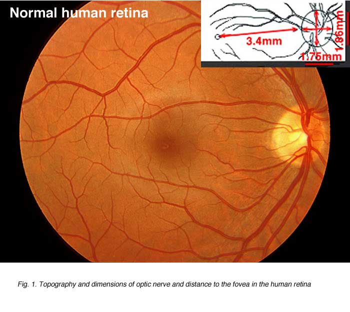
2. Size of optic nerve head or disc.
1.86 x 1.75 mm
3. Degrees and distance in micometers.
- One degree of visual angle is equal to 288 µm on the retina without correction for shrinkage (Drasdo and Fowler (1974).
4. Foveal position.
- 11.8o or or 3.4 mm temporal to the optic disk edge
5. Cross diameter of the macula.
- 3 mm of intense pigmentation, surrounded by 1 mm wide zone of less pigmentation (Polyak, 1941).
6. Cross diameter of the central fovea from foveal rim to foveal rim.
- 1.5 mm (Polyak, 1941)
- 1.2-1.5 mm (Ahnelt and Kolb, unpublished data)

7. Cross diameter of central rod free área.
- 400-600 µm (Polyak, 1941)
- 750 µm (Hendrickson and Youdelis, 1984)
- 570 µm (Yamada, 1969)
- 250 µm (Ahnelt et al., 1987)
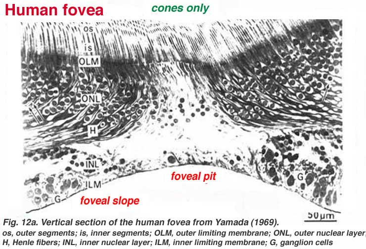
8. Vertical thickness of the fovea from ILM to ELM.
- In the foveal pit 150 µm (Yamada, 1969)
- foveal rim 300 µm
9. Length of foveal axons (Henle fibers).
- 150-300 µm (Ahnelt and Pflug, 1986).
10. Vertical thickness of the retina in different áreas.
- The vertical extent of the retina across the horizontal meridian at different eccentricities is shown in Figure 3. This is taken from data given by Sigelman and Ozanics (1982). The small black numbers are the originals from Sigelman and Ozanics which were measured in typical histological preparations where there is a great deal of shrinkage. The figures in red are those recently measured by Ahnelt (personal communication) in well fixed EM quality material where there is little or no shrinkage. Hence the latter numbers are larger. The numbers are in mm

11. Age when fovea is fully developed.
- Not before 4 years of age (Hendrickson and Youdelis, 1984).
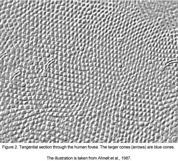
12. Highest density of cones at center of the fovea (counted in a 50 x 50 µm square).
- 147,000/mm2 (Osterberg, 1935)
- 96,900-281,000/mm2 mean161,900/mm2 (Curcio et al., 1987).
- 178,000-238,000/mm2 (Ahnelt et al., 1987).
13. Total number of cones in fovea.
- Approximately 200,000. There are 17,500 cones/degree2. Rod free area is approximately 1o thus there are 17,500 cones in the central rod-free fovea.
14. Total number of cones in the retina.
- 6,400,000 (Osterberg, 1935).
15. Total number of rods in the retina.
- 110,000,000 to 125,000,000 (Osterberg, 1935).
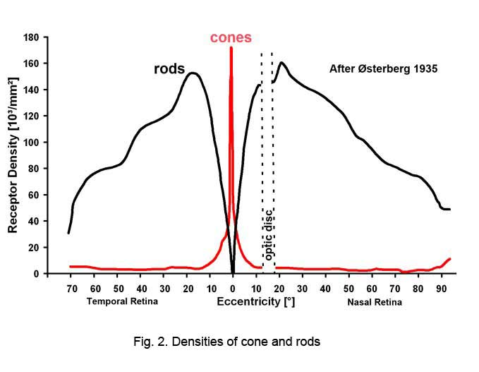
16. Rod distribution.
- Rods peak in density 18o or 5mm out from the center of the fovea in a ring around the fovea at 160,000 rods/mm2. (Fig. 5)
- No rods in central 200 µm.
- Average 80-100,000 rods/mm2
- Rod acuity peak is at 5.2o or 1.5 mm from foveal center where there are 100,000 rods/mm2 (Mariani et al.,1984).
17. Number of axons in the optic nerve.
- 564,776-1,140,030 (Bruesch and Arey, 1942)
- 800,000-1,000,000 (Polyak, 1941)
- 1,200,000 (Quigley et al., 1982; Balaszi et al., 1984).
18. Number of cones to ganglion cells in the fovea.
- 1 cone to 2 ganglion cells out to about 2.2o (Schein, 1988).
19. Number of cones/retinal pigment epithelial cell (RPE).
- 30 cones/RPE in fovea (Rapaport et al., 1995).
20. Number of rods/retinal epithelial cell (RPE).
- In periphery 22 rods/RPE cell
- In rod peak (4-5 mm from foveal center) 28 rods/RPE cell (Rapaport et al.,1995).
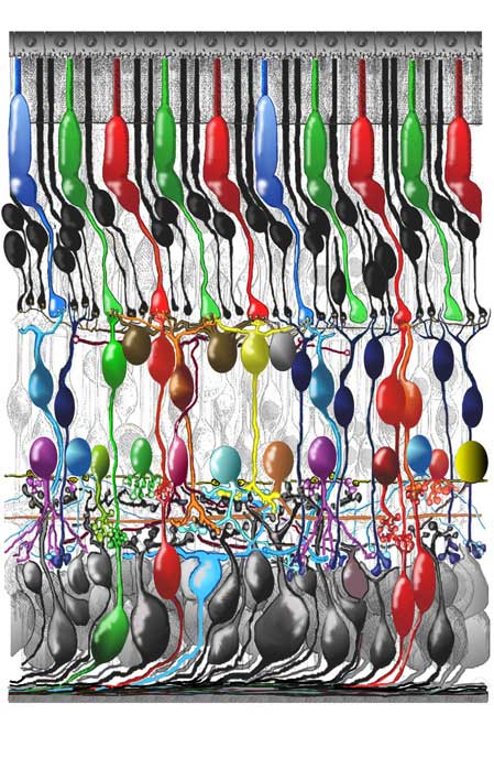
21. Number of neural and glial types in the retina .
The retina consists of many millions of cell types packed together in a tightly knit network spread over the surface of the back of the eye fundus as a thin film of tissue only 1/2 millimeter thick. The retina is like a three layered cake with three layers containing cell bodies of neurons and two filling layers where synapses betwen the neurons occur. There are two basic kinds of photoreceptors, rods and cones. The cones are further subdivided into two types (long and short wavelength sensitive) in the majority of mammals, i.e. most mamals are dichromats and have divariant color vision. In primates a third wavelength sensitive cone has developed closely related to the long wavelength cone type but a little more sensitive in the middle wavelength (i.e. green cone). Thus primates including man are trichromats and have trivariant color vision. Many reptiles, birds and fish have 4 or even 5 types of cone each sensitive to a slightly different peak wavelength. The second order neurons postsynaptic to the photorecepors in the first synaptic (filling layer) (outer plexiform layer) are bipolar cells and horizontal cells. There are 9 types of bipolar cell and 2 to 4 types of horizontal cell in species from mammals to fish. The third order neurons are amacrine cells and ganglion cells that synapse in the inner synaptic filling layer (inner plexiform layer). There are two types of interplexiform cell stretching between both plexiform layers, in most vertebrate retinas.There are approximately 22 types of amacrine cell and 20 types of ganglion cell in the typical mammalian retina. There may be 30 or more amacrine cell types in fish and reptilian retinas and 22 or so ganglion cell types. The increased number of third order neurons is due to the greater information processing taking place in the non mammalian retinas that in mammalian. All vertebrate retinas also contain large numbers of glial cells. The radial Muller cells strech from outer to inner limiting membranes and surround and isolate all neural cell types from each other except at synapses. Microglia arise in times of injury and are blood borne cell types. Astrocytes surround ganglion cell axons and inner retinal blood vessels.Figure 6 shows a drawing of the human retina close to the fovea where all the cell types that have been studied in detail are depicted in their intricate and marvellous network.
22. Useful Units in Vision Science (Wandell, 1995).
Radiometric units represent a physical measurement e.g., radiance is measured in watts sr -1 m-2.
Calorimetric units adjust radiometric units for visual wavelength sensitivity e.g., luminance is measured in candela per square meter, cd/m2.
Lux are units of illumination. Thus a light intensity of 1 candela produces an illumination of 1 lux at 1 meter.
- Scotopic luminance units are proportional to the number of photons absorbed by rod photoreceptors to give a criterion psychophysical result.
- Photopic luminance units are proportional to a weighted sum of the photons absorbed by L- and M-cones to give a criterion psychophysical result.
Typical ambient luminance levels (cd/m2):.
- Starlight: 0.001
- Moonlight: 0.1
- Indoor lighting: 100
- Sunlight: 10.000
- Maximum intensity of common CRT monitors: 100
One Troland (Td) of retinal illumination is produced when an eye with a pupil size of 1 mm2 looks at a surface whose luminance is 1 cd/m2.
Lens focal length: f(meters); lens power= 1/f (diopters).
23. Image formation (Wandell, 1995).
The eyes are 6 cm apart and halfway down the head.
Visual angle of common objects (degrees, deg)
- The sun or moon = 0.5 deg
- Thumbnail (at arm’s length) = 1.5 deg
- Fist (at arm’s length) = 8-10 deg
Visual field (measured from central fixation)
- Monocular: 160 deg (w) x 175 deg (h)
- Binocular: 200 deg (w) x 135 deg (h)
- Region of binocular overlap: 120 deg (w) x 135 deg (h)
Range of pupil diameters: 1-8 mm.
Refractive indices.
- Air: 1.000
- Glass: 1.520
- Water: 1.333
- Cornea: 1.376
Optical power (diopters).
- Cornea: 43
- Lens (relaxed): 20
- Whole eye: 60
- Change in power due to accomodation: 8
Axial chromatic aberration over the visible spectrum: 2 diopters.
Visible spectrum: 370-730 nanometers (nm)
Peak wavelength sensitivity:
- Scotopic: 507 nm
- Photopic: 555 nm
Spectral equilibrium hues:
- Blue: 475 nm
- Green: 500 nm
- Yellow: 575 nm
- No spectral equilibrium: red
References.
- Ahnelt PK, Pflug R. Telodendrial contacts between foveolar cone pedicles in the human retina. Experientia. 1986;42:298–300. [PubMed]
- Ahnelt PK, Kolb H, Pflug R. Identification of a subtype of cone photoreceptor, likely to be blue sensitive, in the human retina. J Comp Neurol. 1987;255:18–34.[PubMed]
- Balazsi AG, Rootman J, Drance SM, Schulzer M, Douglas GR. The effect of age on the nerve fiber population of the human optic nerve. Am J Ophthalmol.1984;97:760–766. [PubMed]
- Bruesch SR, Arey LB. The number of myelinated and unmyelinated fibres in the optic nerve of vertebrates. J Comp Neurol. 1942;77:631.
- Curcio CA, Sloan KR, Packer O, Hendrickson AE, Kalina RE. Distribution of cones in human and monkey retina: individual variability and radial asymmetry.Science. 1987;236:579–582. [PubMed]
- Drasdo N, Fowler CW. Non-linear projection of the retinal image in a wide-angle schematic eye. Br J Ophthalmol. 1974;58:709–714. [PubMed] [Free Full text in PMC]
- Hageman GS, Johnson LV. The photoreceptor-retinal pigmented epithelial interface. In: Heckenlively JR, Arden GB, editors. Principles and practice of clinical electrophysiology of vision. St. Louis: Mosby Year Book; 1991.
- Hendrickson AE, Youdelis C. The morphological development of the human fovea. Ophthalmology. 1984;91:603–612. [PubMed]
- Mariani AP, Kolb H, Nelson R. Dopamine-containing amacrine cells of rhesus monkey retina parallel rods in spatial distribution. Brain Res. 1984;322:1–7.[PubMed]
- Michels RG, Wilkinson CP, Rice TA. (). Retinal detachment.The C.V. Mosby Company.p 17. 1990
- Osterberg G. Topography of the layer of rods and cones in the human retina. Acta Ophthalmol Suppl. 1935;6:1–103.
- Penkhus J. The ora serrata and its anatomical variations. M.S. Thesis. University of California, Los Angeles. 1965
- Polyak SL. The retina. Chicago: University of Chicago Press. 1941
- Quigley HA, Addicks EM, Green WR. Optic nerve damage inhuman glaucoma. III. Quantitative correlation of nerve fibre loss and visual defect in glaucoma ischemic neuropathy and toxic neuropathy. Arch Ophthalmol. 1982;100:135. [PubMed]
- Rapaport DH, Rakic P, Yasamura D, LaVail MM. Genesis of the retinal pigment epithelium in the macaque monkey. J Comp Neurol. 1995;363:359–376. [PubMed]
- Schein SJ. Anatomy of macaque fovea and spatial densities of neurons in foveal representation. J Comp Neurol. 1988;269:479–505. [PubMed]
- Sigelman J, Ozanics V. In: Jokobiec FA, editor. Ocular anatomy, embryology and teratology. Philadelphia: Harper and Rowe; 1982.
- Van Buren JM. The retinal ganglion cell layer. Springfield (IL): Charles C. Thomas. 1963
- Wandell BA. Foundations of vision. Sunderland (MA): Sinauer Associates, Inc. 1965
- Yamada E. Some structural features of the fovea central is in the human retina. Arch Ophthalmol. 1969;82:151–159. [PubMed]